Research Article - Journal of Clinical Dentistry and Oral Health (2022) Volume 6, Issue 4
Assessing the reason for retreatment in anterior teeth - A retrospective study
Ushanthika T and Surendar S*
Department of Conservative Dentistry and Endodontics, Saveetha Dental College, Saveetha University, Saveetha Institute of Medical and Technical Sciences, Chennai, Tamil Nadu, India
- Corresponding Author:
- Surendar S
Department of Conservative Dentistry and Endodontics
Saveetha Dental College, Saveetha University
Saveetha Institute of Medical and Technical Sciences
Chennai, Tamil Nadu, India
E-mail: surendars.sdc@saveetha.com
Received: 27-Jun-2022, Manuscript No. AACDOH-22-68002; Editor assigned: 29-Jun-2022, PreQC No. AACDOH-22-68002(PQ); Reviewed: 14-Jul-2022, QC No.AACDOH-22-68002; Revised: 19-Jul-2022, Manuscript No. AACDOH-22-68002(R); Published: 26-Jul-2022, DOI:10.35841/aacdoh-6.4.119
Citation: Ushanthika T, Surendar S. Assessing the reason for retreatment in anterior teeth - A retrospective study. J Clin Dent Oral Health. 2022;6(4):119
Abstract
Introduction: The endodontic re-treatment demand is increased, because the observations of numerous cross-sectional studies showed that an increased percentage of root filled teeth have evidence of apical periodontitis radiographically and other major reasons that can lead to re-treatment. Aim: The aim of the present study was to assess the reason for retreatment of endodontically treated teeth. Materials and Methods: A Retrospective analysis of all the cases who underwent retreatment was taken and the reasons were assessed. The data was retrieved among the overall data of patients visiting Saveetha Dental College from June 2019-March 2020. The data for 210 patients who underwent retreatment was entered in Excel Spreadsheets and the collected data was analysed using SPSS software version 19. Chi square test was used to statistically evaluate the results. Results: The results of this study show that the main cause of retreatment is incomplete obturation (53.81%), (28.57%) of retreatment is caused due to lesion and periapical pathology, (3.33%) is due missed canals, (10.95%) is due to symptomatic RCT and 3.33% is due to other reasons. The chi-square test was done between age and reason for retreatment and was found to be significant (p value=0.001<0.05; statistically significant). Conclusion Within the limitations of the current study, in this study the main reason for endodontic retreatment were incomplete root canals obturation followed by lesions.
Keywords
Retreatment, Reason, Lesion, Anterior teeth, Innovative.
Introduction
Studies have shown that success rates of root canal therapy generally approach 90 percent [1]. When treatment fails, conventional endodontic retreatment has been suggested as preferable to surgical intervention [2]. Endodontic retreatment has been defined as a procedure performed on a tooth that has received prior attempted definitive treatment resulting in a condition requiring further endodontic treatment to achieve a successful result [3]. According to Bergenholtz et al. [3,4] retreatment usually results in successful outcomes. Endodontic failures can be attributable to inadequacies in shaping, cleaning and obturation, iatrogenic events, or reinfection of the root canal system, when the coronal seal is lost after completion of root canal treatment. The literature shows that many factors are considered responsible for endodontic treatment failure. These include residual necrotic pulp tissue, presence of periradicular infection, periodontal disease, root fractures, broken instruments, mechanical perforations, root canal over fillings, root canal underfillings, missed canals or unfilled canals [5]. Regardless of the aetiology, the sum of all causes is leakage and bacterial contamination. These endodontic failures can be managed by two ways either extraction of the teeth or retreatment [6].
The failure to localize and treat all of the canals of the root canal systems on the part of the operator is considered as one of the major causes of the root canal treatment failures. It has been shown that in majority of cases the general dental practitioners were responsible for the endodontic failures.
The risk of missing anatomy is enhanced due to the intricacy of the root canal system. All the teeth may be found with extra roots/ or canals, but the incidence of this observation is maximum in premolars and molars [6,7].
The standard of coronal restoration has an effect on the periapical status of the root filled teeth [8]. The outcome of a poor root canal filling can be favorable, if the quality of coronal restoration is good. On the other hand a tooth with poor coronal restoration, but having a well cleaned, prepared and well obturatred root canal system may fail shortly [9]. The endodontic retreatment demand is increased, because the observations of numerous cross-sectional studies showed that an increased percentage of root filled teeth have evidence of apical periodontitis radiographically. One of the most influential factors affecting the prognosis of endodontic treatment is the preoperative condition of the tooth. If the tooth has a preoperative peri-apical radiolucent lesion, then it may have a lower success rate up to 20% than the tooth without such preoperative periapical radiolucent lesion [9,10].
Retreatment procedures can be performed at least in all cases with persisting pain, the presence of clinical signs such as swelling or sinus tract, and in teeth with periapical pathosis refractory to endodontic therapy [11]. However, differences in treatment planning choices do exist and are dependent on educational background, clinical experiences, attitudes and values of involved persons, and also economic resources [12].
There are many reasons for the wide variety of outcomes. Several aspects can be attributed to the way in which endodontic successes and failures are reported. Some important factors are the frequency of recall evaluations, operator’s ability, tooth selection, number of cases evaluated, patient’s subjective response to and compliance with treatment, method of determining failures, and subjective interpretation of the results. Endodontic failures are associated most often with periapical pathosis and pain. The decision to perform nonsurgical conventional retreatment, microsurgical endodontics, or even extraction and placement of an implant must be assessed carefully. There have been considerable improvements in endodontic microsurgery techniques that allow for the once-hopeless tooth to be salvaged [13]. These techniques and procedures are still limited by the amount of pulp tissue, bacteria, and any other irritants that can be removed successfully [13,14]. Therefore, a diligent examination of the suspected tooth must be performed to gather information so that the proper treatment can be rendered. Our team has extensive knowledge and research experience that has translated into high quality publications [15-34]. The aim of the study is to assess the reason for retreatment in anterior teeth.
Materials and Methods
A retrospective study was carried out among young adults reporting to Saveetha Dental College and Hospital. The study was conducted between November 2020- August 2021. The study population consisted of 210 patients who needed endodontic retreatment. Ethical approval was obtained from the Institutional Ethical Committee and Scientific Review Board (SRB) of Saveetha Dental College. The data were collected by analyzing the records of 86,000 patients between November 2020-August 2021. The data consisted of 848 patients who required endodontic retreatment for anterior teeth. The data includes the patient's details, the reason for retreatment and tooth number of endodontic failure. The tooth was assessed radiographically. Variables such as age, gender, tooth number and reason for retreatment were recorded. Incomplete, censored and repeated data were excluded from the study.
Statistical analysis
The Data analysis was done by collecting data and was entered in an Excel sheet and subjected to statistical analysis using SPSS software. Chi square tests were done between the gender, age, tooth region and the reason for retreatment of endodontically treated teeth. The independent variables were patient name and PID number while dependent variables are cause of retreatment, age and gender. The level of significance is p<0.05.
Results
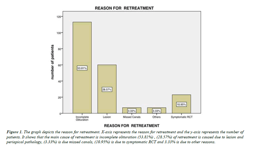
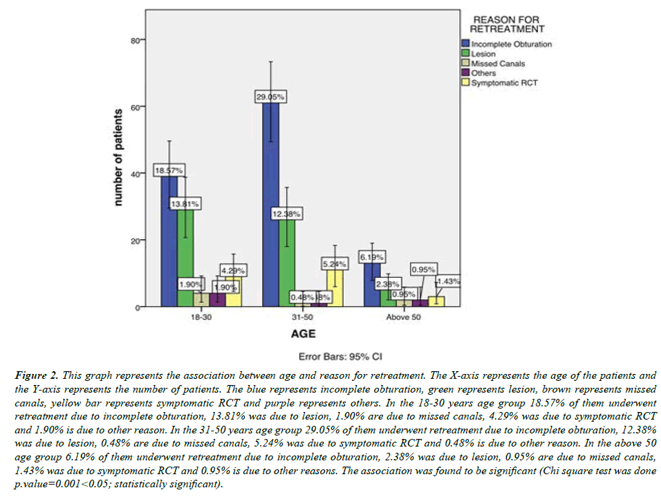
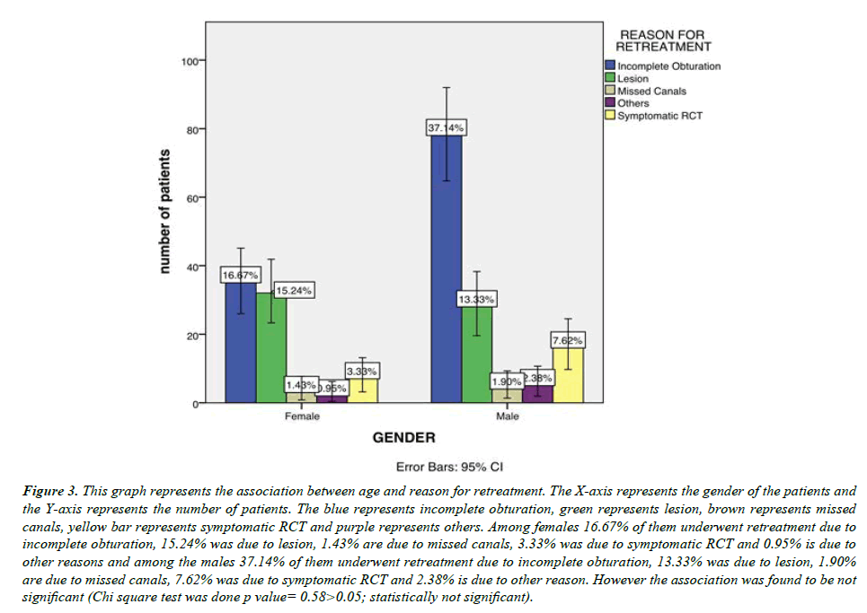
In this study, the patients in this study 38% were females and 62% were males. The age group of the patients were in the 18-30 category there were 41%, 31-50 age group there were 47% and in the above 50 category there were 12% patients. According the data obtained this study shows that the main cause of retreatment is incomplete obturation [53.81%] , [28.57%] of retreatment is caused due to lesion and periapical pathology, [3.33%] is due missed canals, [10.95%] is due to symptomatic RCT and 3.33% is due to other reasons (Figure 1). The most common tooth which underwent retreatment was 11 and 21. Association between age group and reason for treatment was displayed as graphs (Figure 2), the 18-30 years age group 18.57% of them underwent retreatment due to incomplete obturation, 13.81% was due to lesion, 1.90% is due to missed canals, 4.29% was due to symptomatic RCT and 1.90% is due to other reason. In the 31-50 years age group 29.05% of them underwent retreatment due to incomplete obturation, 12.38% was due to lesion, 0.48% is due to missed canals, 5.24% was due to symptomatic RCT and 0.48% is due to other reason. In the above 50 age group 6.19% of them underwent retreatment due to incomplete obturation, 2.38% was due to lesion, 0.95% are due to missed canals, 1.43% was due to symptomatic RCT and 0.95% is due to other reasons. It was found to be significant, the, most common age group to undergo retreatment was the 31-50 age group. Gender and reason for retreatment was associated (Figure 3), Among females 16.67% of them underwent retreatment due to incomplete obturation, 15.24% was due to lesion, 1.43% are due to missed canals, 3.33% was due to symptomatic RCT and 0.95% is due to other reasons and among the males 37.14% of them underwent retreatment due to incomplete obturation, 13.33% was due to lesion, 1.90% are due to missed canals, 7.62% was due to symptomatic RCT and 2.38% is due to other reason. However the association was found to be not significant. Hence, there was no significant relation between gender and reason for retreatment.
Figure 1: The graph depicts the reason for retreatment. X-axis represents the reason for retreatment and the y-axis represents the number of patients. It shows that the main cause of retreatment is incomplete obturation (53.81%) , (28.57%) of retreatment is caused due to lesion and periapical pathology, (3.33%) is due missed canals, (10.95%) is due to symptomatic RCT and 3.33% is due to other reasons.
Figure 2: This graph represents the association between age and reason for retreatment. The X-axis represents the age of the patients and the Y-axis represents the number of patients. The blue represents incomplete obturation, green represents lesion, brown represents missed canals, yellow bar represents symptomatic RCT and purple represents others. In the 18-30 years age group 18.57% of them underwent retreatment due to incomplete obturation, 13.81% was due to lesion, 1.90% are due to missed canals, 4.29% was due to symptomatic RCT and 1.90% is due to other reason. In the 31-50 years age group 29.05% of them underwent retreatment due to incomplete obturation, 12.38% was due to lesion, 0.48% are due to missed canals, 5.24% was due to symptomatic RCT and 0.48% is due to other reason. In the above 50 age group 6.19% of them underwent retreatment due to incomplete obturation, 2.38% was due to lesion, 0.95% are due to missed canals, 1.43% was due to symptomatic RCT and 0.95% is due to other reasons. The association was found to be significant (Chi square test was done p.value=0.001<0.05; statistically significant).
Figure 3: This graph represents the association between age and reason for retreatment. The X-axis represents the gender of the patients and the Y-axis represents the number of patients. The blue represents incomplete obturation, green represents lesion, brown represents missed canals, yellow bar represents symptomatic RCT and purple represents others. Among females 16.67% of them underwent retreatment due to incomplete obturation, 15.24% was due to lesion, 1.43% are due to missed canals, 3.33% was due to symptomatic RCT and 0.95% is due to other reasons and among the males 37.14% of them underwent retreatment due to incomplete obturation, 13.33% was due to lesion, 1.90% are due to missed canals, 7.62% was due to symptomatic RCT and 2.38% is due to other reason. However the association was found to be not significant (Chi square test was done p value= 0.58>0.05; statistically not significant).
Discussion
The failure of endodontic treatment occurs, if the treatment has not been done up to the acceptable standards. The major factors responsible for endodontic treatment failure are the persistent microbial infection in the root canal system and periradicular tissue. In the present study the most common factors observed, responsible for endodontic treatment failure were incomplete root canals obturation followed by lesion. It shows that the main cause of retreatment is incomplete obturation 53.81% and 28.57% of retreatment is caused due to lesion and periapical pathology. According to the Washington study (59%) [35] Cause of retreatment was due to Incomplete obturation and (50%) in the Petersson, et al. study [36], and poor obturation quality noted in this investigation (65%) were all high percentage negative influencing factors. Inadequate obturation is likely associated with difficulty in or incomplete instrumentation of the canal system. The lack of appropriate canal shaping only increases the clinical difficulty of subsequent cleaning and obturation procedures.
Among the teeth in the anterior region the central incisors were more prone to retreatment. The similar findings from the other similar studies, showing that the quality of the root canal filling has an influence on the prognosis of endodontic treatment, support the findings of the present study [36,37].
In our study retreatment was most seen in the 31-50 years age group 29.05% of them underwent retreatment due to incomplete obturation, 12.38% was due to lesion, 0.48% is due to missed canals, 5.24% was due to symptomatic RCT and 0.48% is due to other reason. Age may be an important factor for the success of a root canal treatment in an individual (Figure 2). No significant association was found in our study with gender (Figure 3). It was found that the majority of the endodontic failures were found in the age group range from 31-50 years. The obvious reason for the high failure rate in the age group III may be the calcified canals in older age groups. Second reason may be the uncooperative behaviour, poor oral hygiene maintenance and low literacy rate.
Conclusion
Within the limitations of the current study, the main reason for endodontic retreatment were incomplete root canal obturation followed by lesion and there was found to be an association between age and reason for retreatment. The reason for retreatment must be assessed for better endodontic treatment outcomes.
Acknowledgement
The authors sincerely acknowledge the faculty Medical record department and Information technology department of SIMATS for their tireless help in sorting out datas pertinent to this study.
Source of Funding
The present project is supported by SOMA Beverages.
Saveetha Dental College and Hospitals, Saveetha Institute of Medical and Technical Science, Saveetha University, India.
Conflict of Interest
The authors declare that there were no conflicts of interest in the present study.
References
- Lewis RD, Block RM. Management of endodontic failures. Oral Surgery, Oral Medicine, J Oral Pathol. 1988;66(6):711-21.
- Allen RK, Newton CW, Brown Jr CE. A statistical analysis of surgical and nonsurgical endodontic retreatment cases. J Endod. 1989;15(6):261-6
- Cohen S, Burns R. Pathways of the pulp 8th ed. St Louis Mosby. 2002;2.
- Bergenholtz G, Lekholm U, Milthon R, et al. Retreatment of endodontic fillings. Eur J Oral Sci. 1979;87(3):217-24.
- Engström B, Frostell G. Experiences of bacteriological root canal control. Acta Odontol Scand. 1964;22(1):43-69.
- Schilder H. Filling root canals in three dimensions. Dent Clin N Am. 1967;11(3):723-44.
- Abou-Rass M. Evaluation and clinical management of previous endodontic therapy. J Prosthet Dent. 1982;47(5):528-34.
- Cantatore G, Berutti E, Castellucci A. Missed anatomy: frequency and clinical impact. Endod Topics. 2006;15(1):3-1.
- Kobayashi C, Suda H. New electronic canal measuring device based on the ratio method. J Endod. 1994;20(3):111-4.
- Weiger R, Hitzler S, Hermle G, et al. Periapical status, quality of root canal fillings and estimated endodontic treatment needs in an urban German population. Dent Traumatol. 1997;13(2):69
- Matsumoto T, Nagai T, Ida K et al. Factors affecting successful prognosis of root canal treatment. J Endod. 1987;13(5):239-42..
- Reit C, HOLLENDER L. Radiographic evaluation of endodontic therapy and the influence of observer variation. Eur J Oral Sci. 1983;91(3):205-12.
- Corr R. Comparison of Non-surgical and Surgical Endodontic Retreatment: A Systematic Review (Doctoral dissertation, Loma Linda University).
- Ruddle CJ. Nonsurgical retreatment. J Endod. 2004;30(12):827-45.
- Muthukrishnan L. Imminent antimicrobial bioink deploying cellulose, alginate, EPS and synthetic polymers for 3D bioprinting of tissue constructs. Carbohydr Polym. 2021;260:117774.
- PradeepKumar AR, Shemesh H, Nivedhitha MS, et al. Diagnosis of vertical root fractures by cone-beam computed tomography in root-filled teeth with confirmation by direct visualization: A systematic review and meta-analysis. J Endod. 2021;47(8):1198-214.
- Chakraborty T, Jamal RF, Battineni G, et al. A Review of Prolonged Post-COVID-19 Symptoms and Their Implications on Dental Management. Int J Environ Res Public Health. 2021;18(10):5131.
- Muthukrishnan L. Nanotechnology for cleaner leather production: a review. Environ Chem Lett. 2021;19(3):2527-49.
- Teja KV, Ramesh S. Is a filled lateral canal–A sign of superiority?. J Dent Sci. 2020;15(4):562.
- Narendran K, Jayalakshmi MN, Sarvanan A, et al. Synthesis, characterization, free radical scavenging and cytotoxic activities of phenylvilangin, a substituted dimer of embelin. ijps [Internet]. 2020; 82 (5).
- Reddy P, Krithikadatta J, Srinivasan V, et al. Dental caries profile and associated risk factors among adolescent school children in an urban South-Indian city. Oral Health Prev Dent. 2020;18(1):379-86.
- Sawant K, Pawar AM, Banga KS, et al Dentinal Microcracks after Root Canal Instrumentation Using Instruments Manufactured with Different NiTi Alloys and the SAF System: A Systematic Review. App Sci. 2021;11(11):4984.
- Bhavikatti SK, Karobari MI, Zainuddin SL, et al. Investigating the antioxidant and cytocompatibility of Mimusops elengi Linn extract over human gingival fibroblast cells. Int J Environ Res Public Health. 2021;18(13):7162.
- Karobari MI, Basheer SN, Sayed FR, et al. An In Vitro Stereomicroscopic Evaluation of Bioactivity between Neo MTA Plus, Pro Root MTA, BIODENTINE & Glass Ionomer Cement Using Dye Penetration Method. Materials. 2021;14(12):3159.
- Rohit Singh T, Ezhilarasan D. Ethanolic extract of Lagerstroemia Speciosa (L.) Pers., induces apoptosis and cell cycle arrest in HepG2 cells. Nutr Cancer. 2020;72(1):146-56.
- Ezhilarasan D. MicroRNA interplay between hepatic stellate cell quiescence and activation. Eur J Pharmacol. 2020;885:173507.
- Romera, A., Peredpaya, S., Shparyk, Y, et al. Bevacizumab biosimilar BEVZ92 versus reference bevacizumab in combination with FOLFOX or FOLFIRI as first-line treatment for metastatic colorectal cancer: a multicentre, open-label, randomised controlled trial. Lancet Gastroenterol & Hepatol. 2018;3(12):845-855.
- Raj R K. β?Sitosterol?assisted silver nanoparticles activates Nrf2 and triggers mitochondrial apoptosis via oxidative stress in human hepatocellular cancer cell line. J Biomed Mater Res Part A. 2020;108(9):1899-908.
- Vijayashree Priyadharsini J. In silico validation of the non?antibiotic drugs acetaminophen and ibuprofen as antibacterial agents against red complex pathogens. J Periodontol. 2019;90(12):1441-8.
- Priyadharsini JV, Girija AS, Paramasivam A. In silico analysis of virulence genes in an emerging dental pathogen A. baumannii and related species. Arch Oral biol. 2018;94:93-8.
- Uma Maheswari TN, Nivedhitha MS, Ramani P. Expression profile of salivary micro RNA-21 and 31 in oral potentially malignant disorders. Braz Oral Res. 2020;34..
- Gudipaneni RK, Alam MK, Patil SR, et al. Measurement of the maximum occlusal bite force and its relation to the caries spectrum of first permanent molars in early permanent dentition. Int J Clin Pediatr Dent. 2020;44(6):423-8.
- Chaturvedula BB, Muthukrishnan A, Bhuvaraghan A, et al. invaginatus: a review and orthodontic implications. Br Dent J. 2021;230(6):345-50.
- Kanniah P, Radhamani J, Chelliah P, et al. Green synthesis of multifaceted silver nanoparticles using the flower extract of Aerva lanata and evaluation of its biological and environmental applications. Chemistry Select. 2020;5(7):2322-31.
- Ilan Rotstein DDS, John I. Ingle DDS. Ingle’s Endodontics. PMPH USA; 2019. 1326 p.
- Petersson K, Lewin B, Hakansson J, et al. Endodontic status and suggested treatment in a population requiring substantial dental care. Dent Traumatol. 1989 ;5(3):153-8
- Block RM, Lewis RD. Management of endodontic failures: rationale for choice of treatment and methodology. Aust Endod J. 1987;13(1):15-8.
Indexed at, Google Scholar, Cross Ref
Indexed at, Google Scholar, Cross Ref
Indexed at, Google Scholar, Cross Ref
Indexed at, Google Scholar, Cross Ref
Indexed at, Google Scholar, Cross Ref
Indexed at, Google Scholar, Cross Ref
Indexed at, Google Scholar, Cross Ref
Indexed at, Google Scholar, Cross Ref
Indexed at, Google Scholar, Cross Ref
Indexed at, Google Scholar, Cross Ref
Indexed at, Google Scholar, Cross Ref
Indexed at, Google Scholar, Cross Ref
Indexed at, Google Scholar, Cross Ref
Indexed at, Google Scholar, Cross Ref
Indexed at, Google Scholar, Cross Ref
Indexed at, Google Scholar, Cross Ref
Indexed at, Google Scholar, Cross Ref
Indexed at, Google Scholar, Cross Ref
Indexed at, Google Scholar, Cross Ref
Indexed at, Google Scholar, Cross Ref
Indexed at, Google Scholar, Cross Ref
Indexed at, Google Scholar, Cross Ref
Indexed at, Google Scholar, Cross Ref.
Indexed at, Google Scholar, Cross Ref
Indexed at, Google Scholar, Cross Ref
Indexed at, Google Scholar, Cross Ref
Indexed at, Google Scholar, Cross Ref
Indexed at, Google Scholar, Cross Ref
Indexed at, Google Scholar, Cross Ref


