Research Article - Biomedical Research (2017) Volume 28, Issue 7
Application of multimodal MRI in the brain diagnosis of patients with depression
Dongxue Qin1, 2, Lin Sha2, Xiang Li2 and Mei Yi2*1Graduate School, Tianjin Medical University, Tianjin, China
2Department of Radiology, the Second Affiliated Hospital of Dalian Medical University, Dalian, Liaoning, China
- *Corresponding Author:
- Mei Yi
Department of Radiology
The Second Affiliated Hospital of Dalian Medical University, Dalian, China
RAccepted on December 1, 2016
Abstract
Objective: This study aims to investigate the application of multimodal Magnetic Resonance Imaging (MRI) in the brain diagnosis of symptomatic Intracranial Atherosclerotic Stenosis (sICAS) patients with depression; results of which may be an evidence for formulating the early treatment and therapeutic schedule of these patients.
Methods: A total of 120 patients who have severe sICAS with depression admitted in our hospital were randomly divided into the control and observation groups, with 60 cases in each group. All patients were subjected to MRI examinations, including routine MRI+MRA, perfusion-weighted imaging, and highresolution MRI, before the treatment. The patients in the control group underwent the routine internal medical treatment. The patients in the observation group were treated with intravascular stent-assisted angioplasty based on routine internal medical treatment. All patients were followed up for 1 year. The incidence of complications, recurrence rate of sICAS with depression, Mean Transit Time (MTT), Cerebral Blood Volume (CBV), time to peak, and Cerebral Blood Flow (CBF) of patients in two groups were compared before the treatment and after the 12 month follow-up. Meanwhile, the correlation between various imaging indexes and therapeutic schedule was analysed.
Results: Compared with the test results before treatment, MTT was decreased significantly in two groups, and CBV and CBF were increased significantly, and the differences were statistically significant (P<0.05). Compared with that in the control group, MTT was decreased significantly in the observation group, CBV and CBF were increased significantly, the recurrence rate of sICAS with depression was decreased significantly, and the differences were statistically significant (P<0.05).
Conclusion: Endovascular stent implantation reduced the recurrence rate of sICAS with depression. The multimodal magnetic resonance perfusion imaging-associated indexes could be used to evaluate the efficacy of stent implantation in sICAS patients with depression.
Keywords
Multimodal, Magnetic resonance, sICAS with depression.
Introduction
Symptomatic Intracranial Atherosclerotic Stenosis (sICAS) is caused by atherosclerosis, resulting in stroke and even depression. sICAS is an important cause of depression, which is particularly prominent in Southeast Asian countries [1,2].
In this study, the features of sICAS in sICAS patients with depression were evaluated through the one-stop non-invasive MRI technique.
The sensitive imaging indexes predicting the efficacy of stent implantation in sICAS patients with depression were preliminarily established to provide the basis for the early treatment and selection of the therapeutic schedule of sICAS patients with depression [3].
Data and Methods
Clinical data
A total of 120 sICAS patients with depression admitted in our hospital from January 2015 to December 2015 were selected as the research subjects. The patients were randomly divided into the control and observation groups, with 60 cases in each group. A total of 32 males and 28 females comprised the control group, with an average age of 61.5 ± 3.8 years old. Meanwhile, 33 males and 27 females comprised the observation group, with an average age of 62 ± 3.7 years old. No significant difference was noted in the sex, age, or other general data between the two groups (P>0.05).
The inclusion criteria were as follows: severe sICAS with depression as confirmed through clinical and DSA diagnosis, vascular stenosis ≥ 75%, and patients understood the research process and voluntarily participated. Meanwhile, the exclusion criteria include having contraindications for Magnetic Resonance Imaging (MRI) examination and endovascular surgery and presence of comorbid diseases affecting cerebral blood supply. All patients enrolled in this study signed the informed consent, and this study was conducted with approval from the ethics committee.
Research plan
This study was a prospective randomized controlled study. According to the order of treatment, the patients were divided into the control and observation groups using the random number table method, with 60 patient cases in each group. Before the treatment, all patients were subjected to MRI examinations, including routine MRI+MRA, Perfusion- Weighted Imaging (PWI), high-resolution MRI. The patients in the control group underwent routine internal medical treatment. The patients in the observation group were treated with intravascular stent angioplasty based on routine internal medical treatment. All patients were followed up for 12 months. The incidence of complications, recurrence rate of sICAS with depression, Mean Transit Time (MTT), Cerebral Blood Volume (CBV), Cerebral Blood Flow (CBF), and Time to Peak (TTP) were compared before and after the 12 month follow-up. Meanwhile, the correlation between various imaging indexes and therapeutic schedule was analysed.
Observation index
The incidence of complications, recurrence rate of sICAS with depression, MTT, CBV, CBF, and TTP.
Instrument
Philips 3.0T magnetic resonance machine, equipped workstation, 16-channel head, and neck nerve vascular coil.
Statistical methods
The data were statistically analysed using SPSS 19.0 statistical software. The measurement data were expressed using x? ± s and compared using t-test. The count data were compared using χ2 test. P<0.05 indicated that the difference was statistically significant. All tests were two-sided test.
Results
Comparison of imaging indexes of magnetic resonance perfusion in the control and observations groups before the treatment and after 12 month follow-up
Compared with results before the treatment, MTT was decreased significantly in two groups, CBV and CBF were increased significantly, and the differences were statistically significant (P<0.05). Compared with the control group, MTT was decreased significantly in the observation group, CBV and CBF were increased significantly, and the differences were statistically significant (P<0.05), as shown in Table 1.
| Group | MTT (s) | CBV (mL/g) | CBF (mL/g.min) | TTP (s) | ||||
|---|---|---|---|---|---|---|---|---|
| Beforea | Follow-upb | Beforea | Follow-upb | Beforea | Follow-upb | Beforea | Follow-upb | |
| Control group (N=60) | 11.1 ± 0.8 | 8.9 ± 1.7 | 65.4 ± 36.9 | 121.7 ± 42.9 | 462.5 ± 108.6 | 612.9 ± 109.7 | 28.2 ± 4.8 | 22.5 ± 5.1 |
| Observation group (N=60) | 11.0 ± 0.6 | 7.9 ± 1.4 | 66.7 ± 38.4 | 148.5 ± 41.3 | 457.8 ± 103.4 | 661.7 ± 113.5 | 27.9 ± 4.7 | 19.8 ± 4.9 |
| t | 0.77 | 3.52 | 0.19 | 3.49 | 0.24 | 2.39 | 0.35 | 2.96 |
| P | 0.44 | 0.001 | 0.85 | 0.001 | 0.81 | 0.02 | 0.73 | 0.004 |
aBefore the treatment; b12-month follow-up.
Table 1. Comparison of imaging indexes of magnetic resonance perfusion in the control and observation groups before the treatment and after 12 month follow-up.
Patient results of multimodal magnetic resonance examination
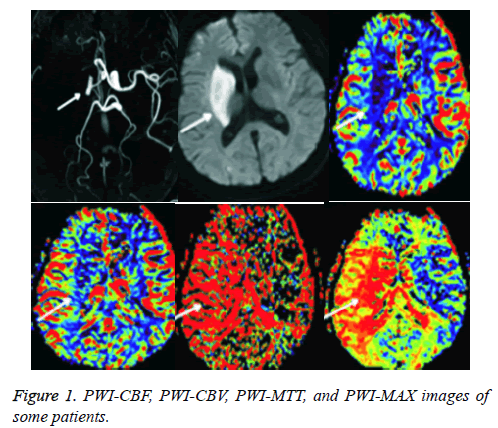
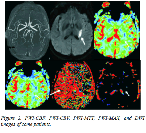
Among 120 cases of sICAS patients with depression, PWICBF and PWI-CBV figures of 81 cases showed low perfusion, 63 cases showed PWI-MTT extension, and 82 cases showed PWI-MAX delay. A total of 87 cases of routine MRI sequences suggested low perfusion, and Diffusion-Weighted Imaging (DWI) suggested infarction lesions, as shown in Figure 1. DWI of 15 patient cases showed significant infarction lesions. No significant perfusion was noted under routine MRI, as shown in Figure 2.
Complications and recurrence rate of sICAS with depression in the control and observation groups after 12 month follow-up
The incidences of complications were similar in the two groups; however, the difference was not statistically significant (P>0.05). Compared with that of the control group, the recurrence rate of sICAS with depression was significantly decreased in the observation group, and the difference was statistically significant (P<0.05), as shown in Table 2.
| Group | Incidence of complications (cases, %) | Recurrence rate of sICAS with depression (cases, %) |
|---|---|---|
| Control group (N=60) | 4 (6.5%) | 5 (8.3%) |
| Observation group (N=60) | 3 (5%) | 13 (21.7%) |
| χ2 | 0.0 | 4.18 |
| P | 1.0 | 0.04 |
Table 2. Incidence of complications and recurrence rate of sICAS with depression in the control and observation groups.
Discussion
Multimodal MR perfusion imaging is widely used in the diagnosis and treatment of sICAS with depression [4]. However, studies on the efficacy evaluation of intracranial artery stent in sICAS patients with depression are limited. In this study, the recurrence rate of sICAS with depression in the observation (endovascular angioplasty) group was significantly lower than that of the control (routine treatment) group. The detection rate of multimodal MRI was higher than that of routine MRI in detecting the recurrence of sICAS with depression. The associated indexes of Magnetic Resonance (MR) perfusion imaging were significantly improved compared with that of before the treatment. The associated index in the observation group was significantly improved than that of the control group after the 12 month follow-up. Meanwhile, the recurrence rate of sICAS with depression was significantly lower in the observation group than that of the control group, which shows that the associated indexes of MR perfusion imaging might be an indicator of the recurrence of sICAS with depression [5,6]. MR PWI is very sensitive to the change of microcirculation perfusion and can accurately reflect the microcirculation state of the brain tissue. The MTT, CBV, and CBF can accurately reflect the hemodynamic status of ischemic areas in real time. Various reasons, including cerebrovascular stenosis, can cause atherosclerosis, resulting in capillary hypoperfusion in ischemic regions and increasing the MTT, CBV, and CBF. Partial cerebral blood vessels have certain compensatory mechanisms, and when activated, these mechanism result in increased CBV to attain brain metabolism in ischemic areas [7]. If the capillary perfusion pressure continues to decrease and exceeds the CBV compensatory limit, the CBF will decrease.
MR perfusion imaging shows the decrease of CBV and increase of MTT in the early stage of sICAS with depression [8]. Therefore, this method can sensitively predict sICAS with depression. In this study, the MTT, CBV, CBF, and TTP of patients in the observation group were significantly better than those of the control group. Also, the recurrence rate of sICAS with depression was significantly lower in the observation group than that of the control group. The results showed that PWI associated indexes could sensitively predict the different treatment effects. Moreover, the use of stent angioplasty in the treatment of sICAS patients with depression was reported earlier in China. A particular study found that balloon dilation was safe and effective in the treatment of sICAS patients with depression. Another study reported a significantly increased incidence of complications in sICAS patients with depression treated through stent implantation. The consensus of Chinese experts on the treatment of sICAS patients with depression is that stent implantation in the treatment of sICAS still needs further studies. This consensus may be based on to the fact that the stent could significantly open the blood vessels, thus, vascular perfusion can be significantly increased for a short period of time [9,10]. Furthermore, the sample size of this study was limited, the follow-up time was short, and the dynamic changes of the related data were absent in the followup period, hence the certain limitations for the research results. The research conclusions need to be evidenced with further clinical trial data.
Conclusion
Multimodal MR perfusion imaging can be used to predict the efficacy of stent implantation in sICAS patients with depression.
References
- Limon-Pacheco JH, Valdovinos-Flores C, Navarrete-Leon RM. Acetaminophen induces the transcription of the antioxidant proteins thioredoxin 1 and glutaredoxin 1 in the brain and liver of Balb/C mice. Lat Am J Pharm 2016; 35: 2029-2035.
- Nitkunan A, Barrick TR, Charlton RA. Multimodal MRI in cerebral small vessel disease: its relationship with cognition and sensitivity to change over time. Stroke 2008; 39: 1999-2005.
- Fang Z, Chen S, Chen Y. Pluronic P85 Enhances the delivery of phenytoin to the brain versus verapamil in vivo. Lat Am J Pharm 2014; 33: 812-818.
- Solliman MA, Hassali MA, Al-Haddad MS. Treatment outcomes of new smear positive pulmonary tuberculosis patients in north east Libya. Lat Am J Pharm 2012; 31: 567-573.
- Hlaihel C, Guilloton L, Guyotat J. Predictive value of multimodality MRI using conventional, perfusion, and spectroscopy MR in anaplastic transformation of low-grade oligodendrogliomas. J Neuro Oncol 2010; 97: 73-80.
- Duarte MR, Dranka ERK, Perez E. Anatomical diagnosis of verbena montevidensis spreng. (verbenaceae) supported by light and scanning electron microscopic images. Lat Am J Pharm 2015; 34: 1476-1480.
- Kuzniecky RI, Bilir E, Gilliam F. Multimodality MRI in mesial temporal sclerosis: relative sensitivity and specificity. Neurology 1997; 49: 774-778.
- Tudorascu DL, Rosano C, Venkatraman VK. Multimodal MRI markers support a model of small vessel ischemia for depressive symptoms in very old adults. Psychiat Res Neuroi 2014; 224: 73-80.
- Iftekharuddin KM, Zheng J, Islam MA. Fractal-based brain tumor detection in multimodal MRI. Appl Math Comput 2009; 207: 23-41.
- Bang O. Multimodal MRI for ischemic stroke: from acute therapy to preventive strategies. J Clin Neurol 2009; 5: 107-119.

