Research Article - Journal of Clinical Dentistry and Oral Health (2022) Volume 6, Issue 1
Analysis of patients undergoing composite restoration in the anterior teeth with fluorosis - A retrospective study
Farhat Yaasmeen Sadique Basha andSurenda Sugumaran*
Department of Conservative Dentistry And Endodontics, Saveetha Dental College, Saveetha Institute of Medical and Technical Sciences, Saveetha University, Chennai, India
- Corresponding Author:
- Surendar Sugumaran
Department of Conservative Dentistry and Endodontics
Saveetha Dental College
Saveetha Institute of Medical and Technical Sciences
Saveetha University, Chennai, India
E-mail: surendars.sdc@saveetha.com
Received: 01-Jan-2022, Manuscript No. AACDOH-22-52396; Editor assigned: 04-Jan-2022, PreQC No. 04-Jan-2022 AACDOH-22-52396 (PQ); Reviewed: 14-Jan-2022, QC No. AACDOH-22-52396; Revised: 21-Jan-2022, Manuscript No. AACDOH-22-52396 (R); Published: 28-Jan-2022, DOI: 10.35841/aacdoh-6.1.104
Citation:Basha FYS, Sugumaran S. Analysis of patients undergoing composite restoration in the anterior teeth with fluorosis - A retrospective study. J Clin Dentistry Oral Health. 2022; 6(1):11-15.
Abstract
Background and aim: Endemic fluorosis has been a public health concern in India for quite some time now. Approximately 62 million people including children suffer from fluorosis due to the consumption of water containing high fluoride concentration. Symptoms of fluorosis may range from white specks or streaks to dark brown stains and pitted enamel. The aim of this study is to determine the prevalence of patients undergoing composite restoration in anterior teeth with fluorosis. Methodology: In this study, the dental records of patients that received composite restoration in their anterior teeth in Saveetha Dental College and Hospitals were screened from July 2019 to February 2021. They were filtered for patients who were diagnosed with fluorosis. The data was extracted from the DIAS. The data was processed in SPSS. Descriptive statistics and Chi-square test was done. The results were then presented as graphs. Results: About 32% of the patients that received anterior restorations while having fluorosis belonged to the age group 21-25 years. Out of which 80% were males. Around 4% of the patients with moderate fluorosis had restorations in the lower arch and 36% of the patients had mild fluorosis and restorations in the upper arch. Conclusion: Within the limits of the study we can conclude that mild fluorosis is more prevalent among the patients and most of the patients with fluorosis received restorations in the upper anteriors. Lower anteriors were only involved in patients with moderate fluorosis.
Keywords
Fluorosis, Water, Fluoride concentration, White specks, Dark stains, Innovative technique, Enamel
Introduction
Fluorosis is one of the major public health problems in India. Approximately 62 million adults out of whom six million are children, suffer from fluorosis due to the consumption of water with high fluoride concentrations [1]. Fluoride is known for its anti-cariogenic properties and hence commercial fluoridated toothpastes, water, salts, etc came into being. Low fluoride intake of less than 1.2-6 ppm results in increased chances of caries. However increased intake of fluoride leads to fluorosis [2,3]. Fluorosis was first reported in a study conducted in 1937 as a public health problem in parts of India [4]. Symptoms of fluorosis may range from just white specks or streaks to dark brown stains and rough-pitted enamel.
Fluoride is strongly electronegative and is attracted to the positively charged calcium present in our teeth and bones. This results in dental and skeletal fluorosis [5]. According to the World Health Organization, the highest prevalence of fluorosis is found in China and India [6]. Another study stated that 10 out of 29 districts in Tamil Nadu have shown evidence of high fluoride content in their water resources [7]. A Few researches have shown that the risk of developing dental fluorosis is higher between the ages of 3 to 6 years because this is when permanent dentition develops [8].
The increased prevalence of dental fluorosis has caused esthetic concerns for many patients. Fluorosis, although not affecting a patient’s physiological functions, was still seen as a defect. This is due to more dental awareness and the desire for an aesthetic dental appearance. Patients have been opting for various dental treatments to treat fluorosis [9,10]. Some of the treatments include restorations, vital bleaching, veneering, etc. A study conducted in 2011 showed that out of the 256 composite restorations placed in the patients, 170 were in the anterior teeth and 86 were in the posterior teeth [11]. This shows that aesthetics plays a major role in patients reporting to clinics for dental treatment and restorations.
Our team has extensive knowledge and research experience that has translated into high quality publications [12-31]. The aim of this study was to evaluate the prevalence of composite restorations in the anterior teeth of patients having fluorosis.
Materials and Method
Study setting
This is a Hospital based retrospective study carried out by reviewing the dental records of patients that reported to Saveetha dental college seeking composite restoration in the anterior teeth between July 2019 to February 2021. Data collection was reliable and with evidence.
Sampling
The patients that reported for composite restorations were screened for those who had fluorosis and received restoration in the anterior teeth. The sample consisted of 50 patients.
Data collection
The patient data was collected from the DIAS. It included parameters like Hospital record number, Name, Gender, Age, Tooth number, Arch and Classification of fluorosis. Data collected was exported into Microsoft Excel 2010.
Data analysis
The data obtained was tabulated and statistically analyzed using Statistical Package for Social Sciences (SPSS version 20.0) for Windows. The descriptive data obtained was plotted in bar graphs. The statistical test used for analysis was the Chi-Square test using SPSS software. Age and Gender were considered as independent variables.
Discussion
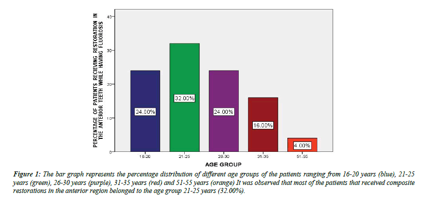
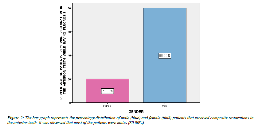
From the graphs we found that most of the patients that received composite restorations in the anterior region belonged to the age group 21-25 years (Figure 1). This can be attributed to the fact that dental awareness and the desire for a good dental appearance has been recently growing in the younger generation. Since the young working class belongs to this age group, we can find most of the patients receiving composite restorations in the anterior teeth in this age group. It was also observed that most of the patients that received composite restorations in the anterior region while having fluorosis were males (Figure 2). A study conducted in Salem revealed that fluorosis was observed in 30.8% children, 31.1% males and 30.3% females [7].
Figure 1: The bar graph represents the percentage distribution of different age groups of the patients ranging from 16-20 years (blue), 21-25 years (green), 26-30 years (purple), 31-35 years (red) and 51-55 years (orange) It was observed that most of the patients that received composite restorations in the anterior region belonged to the age group 21-25 years (32.00%).
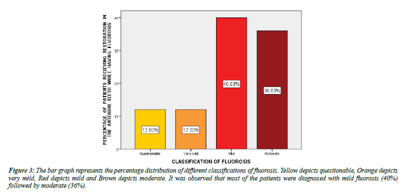
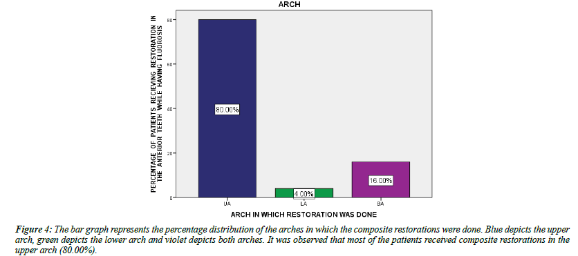
The graphs revealed that most of the patients were diagnosed with mild fluorosis (40%) followed by moderate fluorosis (Figure 3). On analyzing the arches in which the anterior restorations were done, it was observed that 80% of the patients received composite restorations in the upper arch (Figure 4). This can be due to the fact that mostly the upper anteriors are exposed when smiling or talking and hence people give more esthetic importance to the upper arch than the lower arch.
Figure 3:The bar graph represents the percentage distribution of different classifications of fluorosis. Yellow depicts questionable, Orange depicts very mild, Red depicts mild and Brown depicts moderate. It was observed that most of the patients were diagnosed with mild fluorosis (40%) followed by moderate (36%).
Figure 4:The bar graph represents the percentage distribution of the arches in which the composite restorations were done. Blue depicts the upper arch, green depicts the lower arch and violet depicts both arches. It was observed that most of the patients received composite restorations in the upper arch (80.00%).
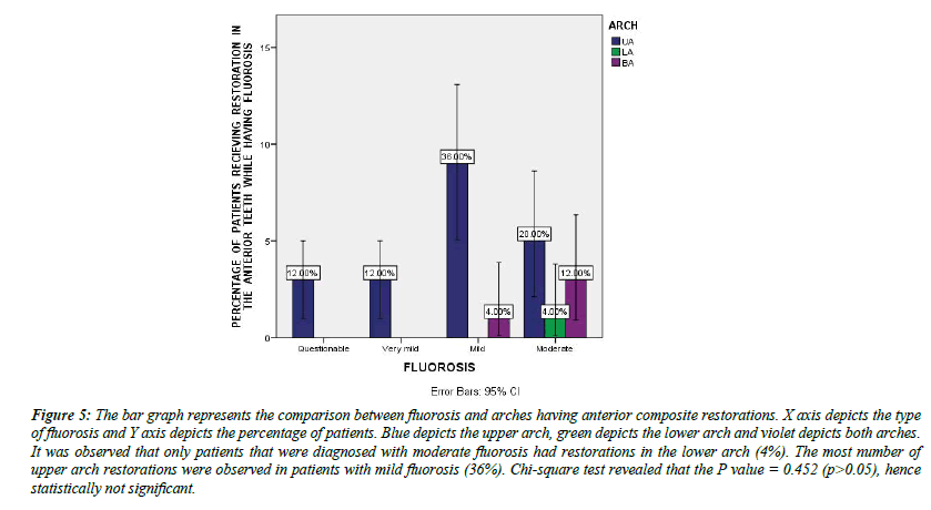
Finally on comparing fluorosis with the arches having anterior restorations we found that only patients that were diagnosed with moderate fluorosis had restorations in the lower arch and the most number of upper arch restorations were observed in patients with mild fluorosis (Figure 5 and Table 1). This shows that as the intensity of the fluorosis increases the patients tend to resort to aesthetic treatments. Not many patients availed treatment for questionable or very mild fluorosis. But in the case of mild fluorosis a large number of patients had reported to the clinic for upper anterior composite restorations. Lower anterior restorations were seen in moderate fluorosis since more staining or discoloration of the teeth will be observed. However the p value was greater than 0.05 hence proving the correlation to be statistically insignificant.
| Fluorosis/ Arch with Restoration | Questionable | Very mild | Mild | Moderate | Total | Level of significance |
|---|---|---|---|---|---|---|
| Upper Anteriors | 3 | 3 | 9 | 5 | 20 | 0.452 |
| Lower Anteriors | 0 | 0 | 0 | 1 | 1 | |
| Both arches | 0 | 0 | 1 | 3 | 4 | |
| Total | 25 | |||||
Table 1: The table displays the correlation between fluorosis and arch having composite restoration.
Figure 5:The bar graph represents the comparison between fluorosis and arches having anterior composite restorations. X axis depicts the type of fluorosis and Y axis depicts the percentage of patients. Blue depicts the upper arch, green depicts the lower arch and violet depicts both arches. It was observed that only patients that were diagnosed with moderate fluorosis had restorations in the lower arch (4%). The most number of upper arch restorations were observed in patients with mild fluorosis (36%). Chi-square test revealed that the P value = 0.452 (p>0.05), hence statistically not significant.
Conclusion
Within the limits of the study we can conclude that mild fluorosis is more prevalent among the patients and most of the patients with fluorosis received restorations in the upper anteriors. Lower anteriors were only involved in patients with moderate fluorosis. Further studies are required on the prevalence of fluorosis, its effect on patients and the aesthetic treatments available.
Acknowledgement
We would like to thank Saveetha Dental College for providing us with this opportunity to conduct a study on the prevalence of patients undergoing composite restoration in the anterior teeth with fluorosis. We would also like to thank our reviewers for their contributions.
Conflicts of Interest
Nil
Source of Funding
The present study is supported by
1. Saveetha Institute of Medical and Technical Sciences
2. Saveetha Dental College and Hospitals
3. Saveetha University
4. Al Shams Energies Private Ltd.
References
- Rao BV, Bharathi MS. Vascular malformations-Treatment modalities. IAIM, 2019; 6(9): 58-65.
- Ghasemi H, Owlia P, Jalali-Nadoushan MR, et al. A clinicopathological approach to sulfur mustard-induced organ complications: a major review. Cutan Ocular Toxicol. 2013;32(4):304-24.
- Buckley D. Congenital Nevi, Melanocytic Naevi (Moles) and Vascular Tumors in Newborns and Children. Prim Care Dermatol 2021;225-231.
- Wagner RS, Gold R, Langer P. Management of capillary hemangiomas. J Pedia Ophthalmol Strabis. 2006;43(6):326.
- Stengler M. Outside the Box Cancer Therapies: Alternative Therapies that Treat and Prevent Cancer. Hay House 2019.
- Mahady K, Thust S, Berkeley R, et al. Vascular anomalies of the head and neck in children. Quant Imag Med Surg. 2015;5(6):886.
- Tyagi I, Syal R, Goyal A. Management of low-flow vascular malformations of upper aero digestive system—role of N-butyl cyanoacrylate in peroperative devascularization. Br J Oral Maxillofac Surg. 2006;44(2):152-6.
- Cooke Macgregor F. Facial disfigurement: problems and management of social interaction and implications for mental health. Aesth Plastic Surg. 1990;14(1):249-57.
- Ianniello A, Laredo J, Loose DA, et al. ISVI-IUA consensus document diagnostic guidelines of vascular anomalies: vascular malformations and hemangiomas. Int Angiol 2015;34:1-2.
- Dhole P, Lohe VK, Sayyad A, et al., A case report on capillary hemangioma and Leukoplakia on Tongue. Med Sci. 2020;24(106):4211-6.
- Pote PG, Banode P, Rawekar S. Lifesaving successful embolization of aggressive vertebral body hemangioma and a large pulmonary arteriovenous malformation. In J Vasc Endovasc Surg. 2021;8(3):269.
- Haile LM, Kamenov K, Briant PS, et al. Hearing loss prevalence and years lived with disability, 1990–2019: findings from the Global Burden of Disease Study 2019. Lancet. 2021;397(10278):996-1009.
- Vos T, Lim SS, Abbafati C, et al. Global burden of 369 diseases and injuries in 204 countries and territories, 1990–2019: a systematic analysis for the Global Burden of Disease Study 2019. Lancet. 2020 ;396(10258):1204-22.
Indexed at, Google Scholar, Cross Ref
Indexed at, Google Scholar, Cross Ref
Indexed at, Google Scholar, Cross Ref
Indexed at, Google Scholar, Cross Ref
Indexed at, Google Scholar, Cross Ref
Indexed at, Google Scholar, Cross Ref
