Image Article - Ophthalmology Case Reports (2021) Volume 5, Issue 1
Acute macular neuroretinopathy as a manifestation of covid-19.
Jack Komro1*, Joseph D Bogaard2, Clinton C Warren2
1 Kirksville College of Osteopathic Medicine, A.T. Still University, BS, USA
2 Medical College of Wisconsin, USA
- Corresponding Author:
- Jack Komro Kirksville College of Osteopathic Medicine A.T. Still University BS, USA E-mail: sa199747@atsu.edu
Accepted date: October 10, 2020
Description
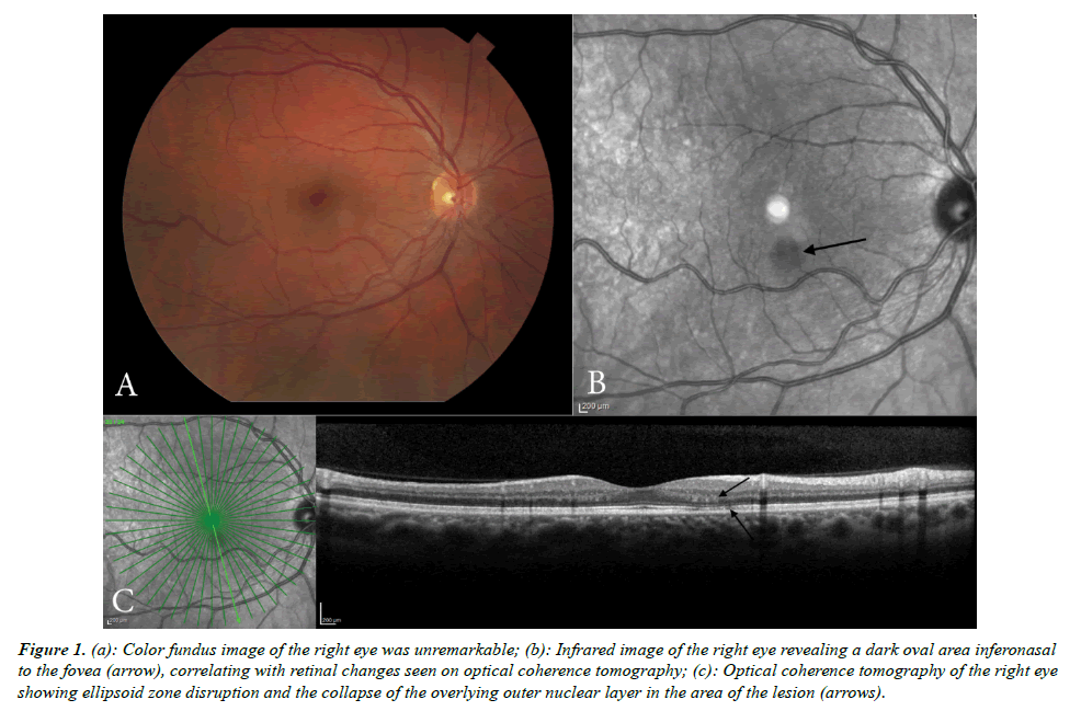
We herein demonstrate a case of a new paracentral scotoma following SARS-CoV-2 infection. A 34-year-old male presented with a two-month history of an acute, oval-shaped paracentral scotoma in the right eye which developed in April 2020, four days following a febrile illness with cough. Nasopharyngeal swab testing confirmed a diagnosis of COVID-19. In June 2020, the patient sought an ophthalmic evaluation for a persistent scotoma. At that time, the patient demonstrated normal visual acuities and an unremarkable funduscopic exam (Figure 1a, right eye only). A superotemporal scotoma was detected on formal visual field testing while infrared imaging (Figure 1b) revealed a dark oval lesion inferonasal to the fovea which correlated with the location of the scotoma. Optical coherence tomography demonstrated an area of ellipsoid zone disruption and collapse of the overlying outer nuclear layer (Figure 1c). Fundus autofluorescent imaging was normal. Flourescein angiography was deferred. These rather subtle findings are suggestive of a late presentation of acute macular neuroretinopathy secondary to COVID-19.
Figure 1: (a): Color fundus image of the right eye was unremarkable; (b): Infrared image of the right eye revealing a dark oval area inferonasal to the fovea (arrow), correlating with retinal changes seen on optical coherence tomography; (c): Optical coherence tomography of the right eye showing ellipsoid zone disruption and the collapse of the overlying outer nuclear layer in the area of the lesion (arrows).
Declaration
The author has no conflict of interest with the material presented in this article.
