Case Report - International Journal of Respiratory Medicine (2021) Volume 6, Issue 1
A typical radiological presentations of COVID-19: Two rare case reports.
Balbir Malhotra, Dilbag Singh, Amritpal Kaur, Harveen Kaur*, Jasvir KaurDepartment of Pulmonary Medicine, Government Medical College, Amritsar Punjab, India
- *Corresponding Author:
- Harveen Kaur
Department of Pulmonary Medicine
Government Medical College
Amritsar, Punjab, India
E-mail: hk_94basra@ymail.com
Accepted on October 20, 2021
Citation: Malhotra B, Singh D, Kaur A, Kaur H, Kaur J. A typical radiological presentations of COVID-19: Two rare case reports. 2021: 6(1): 1-2
Abstract
COVID-19 (Coronavirus Disease 2019) is an infectious disease caused by Severe Acute Respiratory Syndrome Coronavirus 2 (SARS-Cov-2). It has virtually affected every territory of the world except few isolated South Pacific island states and Antarctica. Though RT-PCR testing for the diagnosis of SARS-Cov-2 is the gold standard but in many settings chest imaging has been considered as a part of diagnostic work-up of patients with suspected or probable COVID-19, patients where RT-PCR is either not available or results are delayed or are virtually negative in patients of strongly suspected COVID-19. Chest Imaging has also been considered to complement clinical evaluation in the management of patients or to judge the severity of disease. CT Chest is also more sensitive than RT PCR which takes significantly larger tissue. The sensitivity of CT Chest is 97% versus RT-PCR (60%). Moreover, especially in the early phases of the COVID-19 infection or in the presence of disease with a low viral load, the RT-PCR test may be negative, but CT may reveal significant findings.
Keywords
Chest Radiographs, Chest X ray, COVID-19
Introduction
Common Chest Radiographs show bilateral consolidation especially in lower lung fields and Ground glass opacities (GGO) with a tendency towards the lung periphery while CT findings are more diagnostic and most common are GGO. Other findings are multi-lobe involvement, bilateral lung infiltrates, consolidation and crazy paving pattern. As the disease stage and severity increase, usually consolidation and/or interstitial thickening (crazy paving pattern) begins to appear within the GGO areas. Besides, while GGO or consolidations are healing, it can be seen as a GGO containing fibrotic band or atelectasis in the lung [1-4].
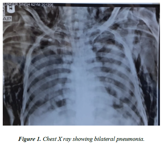
But here we are describing two patients who had uncommon presentations on chest imaging. One patient had Miliary mottling and the other patient had bilateral consolidation with surgical emphysema. These radiological findings are very rare in COVID-19 Figure 1.
Conclusion and Discussion
HS, 62 years old male presented with history of severe breathlessness and fever with Chest X ray showing bilateral pneumonia. Vitals of patient were SPO2 85% on room air, pulse 110 per minute, respiratory rate 32 per minute and blood pressure 150/90 with no other comorbidity. He tested positive on Rapid antigen testing and RT PCR testing for COVID-19. Patient was given symptomatic treatment along with oxygen by high flow nasal cannula, with SPO2 maintained to 92%. 4 days later, patient suddenly became dyspneic, SPO2 dropped to 80% on high flow nasal cannula. Chest X ray done showed bilateral pneumonia with subcutaneous emphysema. Clinically, bilateral crepitations were present. Patient was treated symptomatically, but the patient could not be revived. This case is an uncommon atypical radiological presentation of COVID-19 Figure 2.
RM, 20 years old male referred to us for starting Anti-tubercular treatment for military mottling. Patient had 3 days history of severe breathlessness and fever. On examination, patient was febrile 101◦F, heart rate was 128 per minute, tachypneic, SPO2 76% on room air. HRCT showed military mottling. Sputum for AFB and CBNAAT was negative. Rapid Antigen Test and RTPCR were positive for COVID-19. This case is an uncommon atypical radiological presentation of COVID-19 as Miliary mottling on chest imaging has not been described in the literature.
References
- Ai T, Yang Z, Hou H, et al. Correlation of Chest CT and RT-PCR testing for Coronavirus Disease 2019 (COVID-19) in China: A report of 1014 Cases. Radiology. 2020;296:E32-E40.
- COVID 19 Use of Chest imaging in COVID-19 - a rapid advice guide WHO - 11 June 2020
- Huang P, Liu T, Huang L, et al. Use of chest CT in combination with negative RT-PCR assay for the 2019 novel coronavirus but high clinical suspicion. Radiology. 2020;295:22.
- Xiong Y, Sun D, Liu Y, et al. Clinical and high-resolution CT features of the COVID-19 infection: Comparison of the initial and follow-up changes. Investigative Radiology. 2020;10

