- Biomedical Research (2011) Volume 22, Issue 3
A Study of Morphological Features of Attachment Area of Costoclavicular Ligament on Clavicle and First Rib in Indians and its Clinical Relevance
Anita Rani1, Jyoti Chopra1, Archana Rani1*, Suniti Raj Mishra2, AK Srivastava1, PK Sharma1 and Rakesh Diwan1
1Department of Anatomy, CSM Medical University, Lucknow, India.
2Department of Medical Education, CSM Medical University, Lucknow, India.
- *Corresponding Author:
- Anita Rani
Department of Anatomy
CSM Medical University
Lucknow, Uttar Pradesh
India
Accepted Date: April 17 2011
Abstract
The present study was conducted on the dried specimens of adult clavicles (118) and first ribs (184) of Indian origin. The various patterns of attachment area of costoclavicular liga-ment on clavicle were depressed and rough (30.97%), flat and rough (28.32%), elevated and smooth (19.47%), elevated and rough (9.73%), flat and smooth (6.19%) and depressed and smooth (1.77%). 3.54% clavicles showed no impression on the ligamentous area. On first rib the corresponding area was elevated and rough (50.54%), elevated and smooth(16.31%) flat and rough (15.22%) and flat and smooth(17.93%).64.29% of ossified costal cartilages showed elevated and rough type of pattern .The smooth, elevated and circumscribed im-pressions were considered as faceted apophysis. It was concluded that the most common type of pattern on clavicle was depressed and rough and corresponding to it the most com-mon pattern on first rib was rough elevation. At times, these normal morphological variants may be misinterpreted as pathological in radiological images.
Keywords
clavicle, first rib, first costal cartilage, costoclavicular ligament, costoclavicular joint, rhomboid fossa.
Introduction
The costoclavicular ligament is one of the most important ligaments of the sternoclavicular region which not only strengthens the sternoclavicular joint from its inferior as-pect but also assists in the movements of pectoral girdle [1,2]. This ligament is prone to injury in the painters, con-struction workers and kayakers [3,4,5]. In sternocosto-clavicular hyperostosis, hyperostotic changes are seen in the medial end of clavicle, first costal cartilage and ad-joining part of sternum which may later involve the costoclavicular ligament [6]. As the subclavian vein trav-els just posterior to the costoclavicular ligament, this ana-tomical area becomes crucial while placing venous cathe-ters [7] and pacemakers [8, 9]. It also acts as a landmark for deciding the length of resection of the medial end of clavicle [10].
Costoclavicular ligament is a strong, dense band which extends from the first costal cartilage and the adjoining part of the first rib to the inferior surface of medial part of the shaft of the clavicle [11]. It is disposed as an inverted, truncated cone, flattened antero- posteriorly. The impres-sion area of the costoclavicular ligament on the clavicle is either described as a rough impression [12] or tuberosity [11] or rough oval depressed area [13] which is also commonly termed as rhomboid fossa. Anthropologists have widely studied this fossa as a marker for age and sex determination [14, 15]. These normal morphological vari-ants of attachment area of costoclavicular ligament on clavicle may be misinterpreted as pathological conditions by an inexperienced observer [16]
On the other hand morphology of the attachment area of costoclavicular ligament on the first rib and its cartilage, which is equally important in interpreting the pathology of sternocostoclavicular area, has not yet been described. This lacuna prompted us to study the various patterns of attachment area of costoclavicular ligament on the dried specimens of first rib and clavicle.
Material and Methods
To study the morphology of the area of attachment of costoclavicular ligament on clavicle and first rib, the bones were obtained from the osteology lab of Anatomy Department of Chhatrapati Shahuji Maharaj Medical University, Lucknow, UP, India. Total 184 adult first ribs were studied, out of which 90 were of right side and 94 of left. 113 adult clavicles were observed carefully for the pattern of impression of costoclavicular ligament. 60 clavicles were of right side and 53 belonged to left. The various patterns of the impression were categorized and their incidence was calculated.
Results
The inferior surface of clavicle near its sternal end pro-vides attachment to costoclavicular ligament. This area was well demarcated from rest of the bone in most of the cases and was invariably oval in outline and showed three distinctive types of surface projection viz. flat (34.51%), elevated (29.21%) and depressed (32.74%).
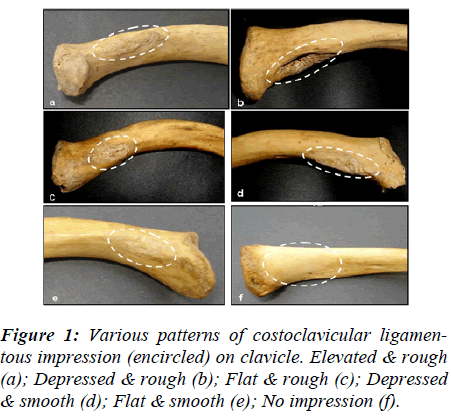
In few clavicles (3.54%) this well circumscribed area was not observed, instead the surface architecture of this par-ticular area was same as that of rest of the inferior surface (Fig 1f) and its incidence was same on both sides. The flat impression was more common on left side where as ele-vated and depressed types of impressions were more common on right side (Table 1).
The surface projections were either rough (69.03%) or smooth (27.43%) and the incidence of roughness as well as smoothness was higher on right side (Table 1b).
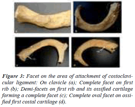
Different combinations of surface projection and architec-ture gave rise to six distinctive patterns of this ligamen-tous impression area on clavicle (Fig 1). These were flat and rough (28.32%), flat and smooth (6.19%), depressed and rough (30.97%), depressed and smooth (1.77%), ele-vated and rough (9.73%) and elevated and smooth (19.47%). The elevated and smooth impressions were circumscribed therefore they were regarded as faceted apophysis (Fig 3a). Except for depressed and smooth and elevated and rough impression all other patterns were more common on right side (Table 1).
The costoclavicular ligament is attached to the inner half of anterior end of first rib, medial to the groove for sub-clavian vein and extends to the adjoining part of first cos-tal cartilage. Out of 184 first ribs, only 30 had ossified costal cartilages. Out of these 30 ossified costal cartilages, 16 were partially ossified and the area of attachment of costoclavicular ligament was unossified and therefore deficient, so we could not comment on the morphology of the ligamentous impression in these cases.
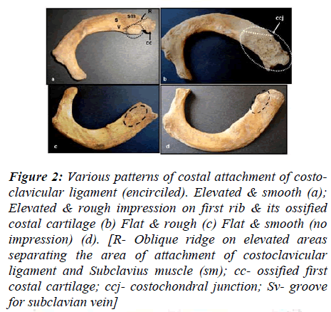
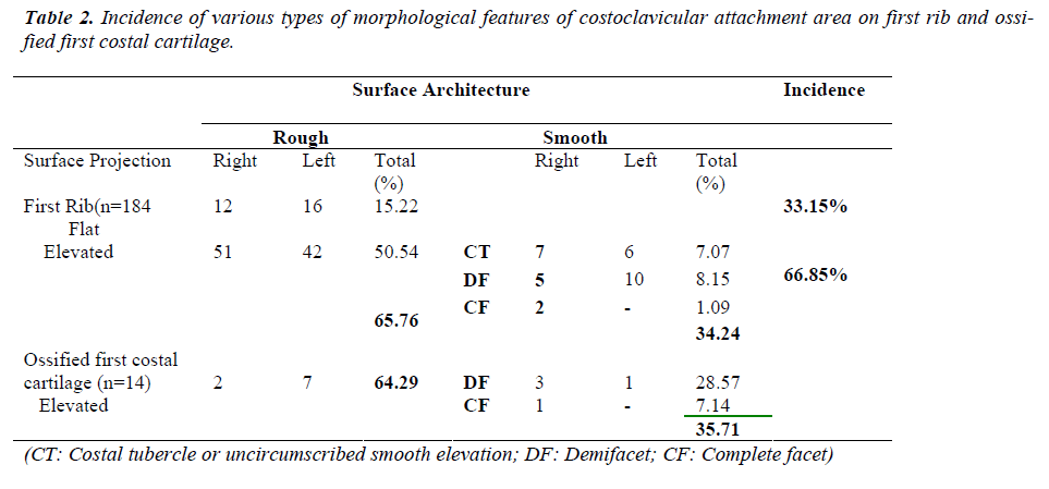
Unlike oval impression of costoclavicular ligament on clavicle, corresponding area in majority of first rib did not exhibit any fix shape or circumscribed margin. In accor-dance to the clavicle, the concerned area on first rib also showed different morphological features. The area medial to the groove for subclavian vein was either elevated (66.85%) or flat (33.15%). Elevation was more common on right side and flatness on the left (Table 2). The ele-vated areas showed a distinct ridge commencing from the inner margin and running forwards and medially, dividing it into postero-medial triangular slope for attachment of costoclavicular ligament and antero-lateral slope for ten-don of subclavius muscle (Fig 2a). It was observed that the surface of none of the first rib was depressed at the site of ligamentous attachment. The surface architecture of the impression area was either rough (65.76%) or smooth (34.24%). Roughness predominated on the right side while smoothness on the left (Table 2).
Figure 2: Various patterns of costal attachment of costo-clavicular ligament (encirciled). Elevated & smooth (a); Elevated & rough impression on first rib & its ossified costal cartilage (b) Flat & rough (c) Flat & smooth (no impression) (d). [R- Oblique ridge on elevated areas separating the area of attachment of costoclavicular ligament and Subclavius muscle (sm); cc- ossified first costal cartilage; ccj- costochondral junction; Sv- groove for subclavian vein]
Different combinations of surface projection and architec-ture observed on first rib were flat and rough (15.22%) (Fig 2c), elevated and rough (50.54%) (Fig 2b) and ele-vated and smooth (16.31%). In 7.07% ribs , this elevated and smooth area was in the form of a tubercle (Fig 2a) while in 8.15% ribs this region showed well circum-scribed area which was half oval and in 1.09% ribs it was oval in shape. These well circumscribed areas were des-ignated as demifacets or facets respectively (Fig 3c, 3b). A fourth type of pattern was also observed in which the concerned area was flat and smooth (17.93%). As in these ribs the surface architecture of the impression area was same as that of rest of the bone so it was considered that in these cases ligament has imparted no impression (Fig 2d). Except for the roughened elevation all other patterns were more common on left side (Table 2).
Out of 14 completely ossified first costal cartilages, 9 car-tilages(64.29%) exhibited elevated and rough (Fig 2b) while 5 (35.71%) showed elevated and smooth impres-sion for costoclavicular ligament. Out of 5 smooth eleva-tions, 4 had demifacets (28.57%) and only one ossified costal cartilage (7.14%) showed presence of well circum-scribed complete facet (Fig.3d). The incidence of facet was higher on right side (Table 2).
Discussion
Due to the recent advancements in the field of imaging techniques, the structural details are easily visualized. A deficient knowledge of normal morphological variants becomes crucial while differentiating between normal and abnormal. The morphology of attachment area of costo-clavicular ligament on the clavicle as well as the first rib is a relatively neglected entity that can potentially cause diagnostic errors.
According to the present study the attachment area of costoclavicular ligament on clavicle was invariably oval in shape and well circumscribed except in a few cases (3.54%), where no impression was seen. Contrary to this, the attachment area on the first rib did not exhibit any well defined margin or shape except in 9.24% cases where oval or half oval impression was present. The shape of this impression on clavicle has unanimously been reported as oval [1,17,13]. No study has been re-ported to the best of our knowledge regarding the shape of ligamentous area over first rib. The size of impression on clavicle was larger as compared to that on first rib and its cartilage which suggests that the shape of the ligament attached to this area is truncated conical, as described classically [13].
We observed three types of surface projection of costo-clavicular attachment area on clavicle i.e. flat, depressed and elevated. There was no significant difference in the incidence of these surface projections. These surface pro-jections were either rough or smooth and the incidence of roughness was much higher. As far as combined charac-ters of ligamentous area were concerned, in the present study, incidence of depressed and rough type of impres-sion was highest (30.97%) followed by flat and rough type, while Cave [1], in his study, on 153 clavicles of European origin, estimated highest incidence of flat and rough type (31%) followed by flat and smooth variety (29%) .The incidence of depressed and rough type of im-pression was only 18% in his study. Flat and smooth type of rhomboid impression was exhibited by a smaller pro-portion of clavicles (6.19%) in our study. The incidence of elevated and rough type of impression was almost same as that observed by Cave [1]. The least common type of impression in the current study was depressed and smooth (1.77%) which in contrast had 10.5% incidence in Cave’s study [1]. Flournoy et al [18], confirmed various skeletal traits produced by costoclavicular ligament on clavicle in North American population and described them as tubercles, roughened impressions, shallow groove like fossae, deep fossae or no impression. However, they did not report the incidence of these patterns.
The most common type of impression found in the pre-sent study ie depressed and rough type is also termed as ‘rhomboid fossa’. Jit & Kaur studied a large population of adult clavicle (789) in Indians and studied exclusively the presence of rhomboid fossa and reported 37.3% incidence which is almost same as that found in the present study [14]. The presence of rhomboid fossa is correlated with the age and sex of the individual by many workers [16,18,19]. Paraskevas et al studied 80 chest radiographs and found 26.88% incidence of excavated type of rhomboid fossa which was correlated with the gender, sidedness and handedness. They found higher incidence of fossa on right side in right handed person and on left side in left handed person so they proposed mechanical theory for the formation of fossa [19].
The corresponding area of attachment of costoclavicular ligament on first rib was either flat or elevated and was uncircumscribed; therefore no shape could be attributed to them. Majority of impressions on first rib as well as on ossified first costal cartilage were of elevated and rough type which were congruent with the most common type of impression on clavicle ie depressed and rough. There was not much difference in the incidence of other three varie-ties of impressions on first rib.
Flat and smooth type of areas on the first rib at the site of attachment of costoclavicular ligament strongly suggests that probably in these cases costoclavicular ligament was either absent or if it was present, its costal attachment was completely shifted to the first costal cartilage. Simultane-ously, clavicular attachment area also exhibited no im-pression in 3.54% cases, again suggesting the absence of costoclavicular ligament in these subjects. In these cases it is presumed that the costoclavicular ligament, instead of being a separate entity, is a part and parcel of capsule of sternoclavicular joint. In early primates also costoclavicu-lar ligament was not a separate entity, but it was continu-ous with sternoclavicular joint capsule [1]. It seems that in these cases the primitive pattern still exists. Higher in-cidence of no impression on first rib as compared to clavicles indicates simply that even when the ligament is present, it is not attached to the first rib instead it is at-tached to its costal cartilage. Our assumption is supported by few authors who have described the attachment of costoclavicular ligament on the first costal cartilage only [12,20].Even absence of costoclavicular ligament has been reported by Tubbs et al [20].
The incidence of demifacets on ossified first costal carti-lage does not correspond with the incidence of demifacets on first rib. This discrepancy might be due to lesser num-ber of available ossified costal cartilages. Presence of fac-eted apophysis on clavicle, first rib and ossified first cos-tal cartilage strongly suggest the existence of a diarthrodial articulation at the attachment area of costo-clavicular ligament. Earliest description of costoclavicular articulation was given by Buchanan, who proposed that in 10% subjects a joint exist between clavicle and first costal cartilage, and in such cases rhomboid ligament forms part of the joint capsule [2]. This fact was further supported by the study of Cave [1]. Though the incidence of facets on first rib ( 9.24%) do not correspond with the incidence of facets on clavicle (19.47%) but this can be justified on the basis of the fact that the costoclavicular joint might exist between clavicle and first costal cartilage only. The com-plete articular facet that we have observed on a single ossified costal cartilage also supports the same. Higher incidence of faceted apophysis at the area of attachment of costoclavicular ligament on clavicle, first rib and first costal cartilage in Indians strongly suggests the develop-ment of a synovial type of costoclavicular joint. Cave proposed that the development of this joint may be secon-dary to wide range of clavicular movements in humans. So, probably it is a next step in the evolution [1].
Most of the authors believe that the costoclavicular liga-ment is made up of anterior and posterior lamina with an intervening constant bursa [1,2,13], while other school of thought denies the bifascicular nature of the same [20,21]. The roughened architecture of the attachment area of costoclavicular ligament on clavicle and first rib, observed in the present study, suggests that there is no intervening bursa and the ligament is attached through out this roughened impression, forming a fibrous articulation between the clavicle and first rib. This articulation be-tween clavicle and first rib by costoclavicular ligament is regarded as fibrous joint by Hollinshed [22].Whereas those clavicles and first ribs where this ligamentous at-tachment area showed smoothness indicates the presence of bursa between the two lamina of costoclavicular liga-ment. In cases where circumscribed elevated and smooth type of impression is present (facets), we hypothesize that this bursa will form the synovial cavity of the diarthrodial articulation between clavicle and first rib. The presence of this synovial variety of costoclavicular joint is also con-firmed in a radiological study by Johnell [23].The prob-able factors responsible for the above morphological variations are environment, genetic constitution, rate and pattern of growth and type of bone remodeling [24,25]
Carrera et al recommended identification of this ligament during surgeries for resection of the clavicle’s proximal extremity [10].But these surgeons should be aware of the fact that this ligament can be either absent or may be merged with the sternoclavicular joint capsule.
The knowledge of various types of ligamentous impres-sion on clavicle and first rib, as observed in the present study, is important clinically because these morphological varieties can be misinterpreted by radiologists while observing sterno-costo-clavicular region. Rhomboid fossa can be misdiagnosed as simple benign fibrous displasia or chronic osteomyelitis [16] whereas elevation on clavicle as well as first rib may be confused with hyperostotic changes. These morphological skeletal traits are also of great value for anthropologists and anatomists so their description should be incorporated in the standard text books of medicine.
Conclusion
The most common type of pattern of costoclavicular liga-mentous attachment area on clavicle is depressed and rough and corresponding to it the most common pattern on first rib is rough elevation. Circumscribed smooth ele-vation on clavicle, first rib and its costal cartilage indi-cates presence of synovial joint among them. The knowl-edge of normal morphological variants of costoclavicular impression areas is clinically useful for radiologists, or-thopedicians, anaesthetists and cardiologists.
References
- Cave AJE. The nature and morphology of the costo- clavicular ligament. J Anat 1961; 95:170-179.
- Wood Jones F. The Bones of The Upper Limb. In Bu- chanan’s Manual of Anatomy. 7th ed. Bailliere Tindall and Cox, London 1946; 268..
- Bearn JG. Direct observations on the function of the capsule of the sternoclavicular joint in clavicular sup- port. J Anat 1967; 101:159-170.
- Booth CM, Roper BA. Chronic dislocation of the ster- noclavicular joint: an operative repair. Clin Orthop Re- lat Res 1979; 140:17-20.
- Robinson CM, Jenkins PJ, Markham PE, Beggs I. Dis- orders of the sternoclavicular joint. J Bone Joint Surg Br 2008; 90(6):685-696.
- Shimizu T, Chigira M. Roentgenological analysis of sternocostoclavicular hyperostosis by computed tomo- graphy. J Jpn Orthop Assn 1989; 63(1):18-24.
- Krutchen AE, Bjarnason H, Stackhouse DJ, Nazarian GK, Magney JE, Hunter DW. The mechanisms of posi- tional dysfunction of subclavian venous catheters. Ra- diology 1996; 200(1):159-163.
- Harada Y, Katsume A, Kimata M, Hikosaka T, Yama- naka S, Akashi K, Hosomi Y, Hirano S, Matsubara H. Placement of pacemaker leads via the extra thoracic subclavian vein guided by fluoroscopy and venography in the oblique projection. Heart Vessels 2005; 20(1):19- 22.
- Magney JE, Flynn DM, Parsons JA, Staplin DH, Chin- Purcell MV, Milstein S, Hunter DW. Anatomical mechanisms explaining damage to pacemaker leads, defibrillator leads, and failure of central venous cathe- ters adjacent to the sternoclavicular joint. Pacing Clin Electrophysiol 1993; 16(3): 445- 457.
- Carrera EF, Archetti Neto N, Carvalho RL, Souza JB, Faloppa F. Resection of the medial end of clavicle: an anatomic study. J Shoulder Elbow Surg 2007; 16(1):112-114
- Schaeffer’s JP. Osteology In: Morris’ Human Anat- omy: A complete systematic treatise. 10th ed. The Blakiston Company, Philladelphia 1942; 187.
- Frazer JE. The Anatomy of The Human Skeleton. 4th ed. J&A Churchill Ltd. London, 1948.
- Williams PL, Bannister LH, Berry MM, Collins P, Dyson M, Dussek JE and Ferguson MWJ. Skeleton System In: Gray’s Anatomy. 38th ed. Churchill Livingston. Edinburgh, 1995; 621.
- Jit I, Kaur H. Rhomboid fossa in the clavicles of North Indians. Am J Phys Anthropol. 1986; 70(1): 97-103.
- Singh J, Singh D. Rhomboid fossa of the clavicle: Is it a reliable estimator of gender and sex of Northwest In dian subjects of Chandigarh zone. J of Punjab Acad Forensic Med & Toxicol 2009; 9(2): 44-46.
- Prado FB, de Mello Santos LS, Caria PHF, Kawaguchi JT, Preza Ad’OG, Daruge E, da Silva RF, Daruge E Incidence of clavicular rhomboid fossa (Impression for costoclavicular ligament) in the Brazilian population: Forensic application. J Forensic Odontostomatol 2009; 27:1:12-16
- Poirier P. La clavicule et ses articulations. J de r Anatomie 1980; 26: 81-103.
- Flournoy LE, Rogers NL, McCormick WF. The rhom- boid fossa of the clavicle as a sex and age estimator. J Forensic Sci 2000; 45 :61-67.
- Paraskevas G, Natsis K, Spanidou S, Tzaveas A, Kitsoulis P, Raikos A, Papaziogas B, Anastasopoulos N. Excavated-type of rhomboid fossa of the clavicle: A radiological study. Folia Morphol (Warsz) 2009; 68(3):163-6.
- Tubbs RS, Loukas M, Slappey JB, McEvoy WC, Lin- ganna S, Shoja MM, Oakes WJ, Surgical and clinical anatomy of the interclavicular ligament, Surg Radiol Anat 2007; 29(5): 357-360.
- Woodburne RT, Burkel WE. Essentials of human anatomy. 8th ed. Oxford University Press, New York, 1988.
- Hollinshed W H. Text Book of Anatomy. 2nd ed. Ox- ford and IBH publishing Co., New Delhi, 1970:170-171
- Johnell IR. The costoclavicular joint. Skeletal Radiol 1986; 15: 25-26
- Humphrey LT, Dean MC, Stringer CB. Morphological variation in great ape and modern human mandibles. J Anat 1999;195: 491-513
- Wood BA, Li Y Willoughby C. Intraspecific variation and sexual dimorphism in cranial and dental variables among higher primates and their bearing on the homi- nid fossil record. J Anat 1991;174: 185-205


