Case Report - Otolaryngology Online Journal (2016) Volume 6, Issue 3
A Multitrabecular Nasal Dermoid Cyst: Case Report
Kiran j shinde and Amit kumar singh*
Pravara institute of medical Sciences, Maharashtra, India
- *Corresponding Author:
- Amit kumar singh
Pravara institute of medical Sciences, Maharashtra, India
E-mail: amit2051india@gmail.com
Received: April 09, 2016; Accepted: May 02, 2016; Published: March 25, 2016
Abstract
Nasal dermoid is a rare developmental anomaly. Unlike other craniofacial dermoid, the nasal lesions can present as a cyst, a sinus or fistula and may have an intracranial extension. We report a case of 11 yr old male child presenting in the ENT OPD of Pravara rural hospital with a tuft of hair over the root of the nose since birth, which is uncommon presentation of nasal dermoid.
Keyword
Nasal dermoid, Congenital lesion, Nasal midline swelling
Introduction
A true dermoid cyst or a dermoid sinus is congenital lesion lined by stratified squamous epithelium, with normal dermal appendages, including-hair follicles, sweat glands, and sebaceous glands. Hair protruding through a punctum is pathognomic for nasal dermoid. The incidence is estimated at 1: 20000 to 1: 40000 births [1]. Nasal lesion account for between 1 and 3 percent of all dermoid and are most common midline nasal mass [2,3]. There is debate on their origin. Theories suggested are 1) failure of the anterior neuropre to close 2) abnormal separation and obliteration of prenasal space 3) inclusion in facial clefts 4) simple inclusion cysts. 5) Aberrant development of skin appendages [4,5]. Both cyst and sinuses may have a connection with an intracranial component through an abnormal foramen caecum or cribriform plate in the anterior cranial fossa [6]. A true dermoid cyst may occur alone subcutaneously on the nasal dorsum superficial to nasal bones without a cutaneous opening. Simple Dermoid cysts appear usually as slowly enlarging mass under the skin of dorsum of nose in any position from tip to glabella with sinus, dorsum hair or intermittent discharge, which over a period of time may simply deform the underlying structures [4]. We are presenting a case report nasal dermoid with protruding hair over dorsum of nose which occur only minority of patients (5%) [7] and it is pathognomic for a nasal dermoid [7].
Case history
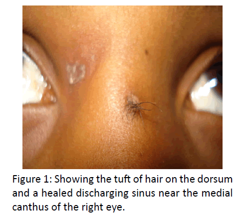
11 yrs old male child was brought to the ENT OPD with complains of a tuft of hair at the root of the nose since birth (Figure 1) and painful swelling with discharge from root of nose since 5 days, which appeared after plucking some hair out by child. The child was otherwise healthy and active and did not have previous history of any medical or surgical illness.
General and systemic examination was normal
Local examination revealed a tuft of hair growing over the root of the nose (just 2 mm lateral to midline on the left) and the skin on the dorsum just lateral to midline on the right was plucked and showed a scar and a sinus within it. There was no active discharge from the sinus. On palpation the underlying tissue was thickened and cord like and showing no fixity to the underlying nasal bone. Blood and urine examination was within normal limits.
MRI was done which showed a small lesion measuring 10 x 3.5 mm with altered signal intensity was seen in prenasal region, posterior to nasal bone and anterior to nasal septum, appearing hyperintensity on T1W, T2W images. The lesion showed low intense signals on STIR images suggesting a fatty component within it. Caudally the lesion was continuous via a narrow tract to the skin surface over nasal bridge in mid sagittal plane distal to the glabella. No obvious close relation of the lesion with the basifrontal gyri in the midline was noted.
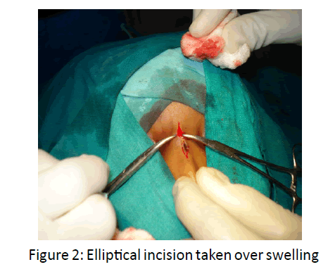
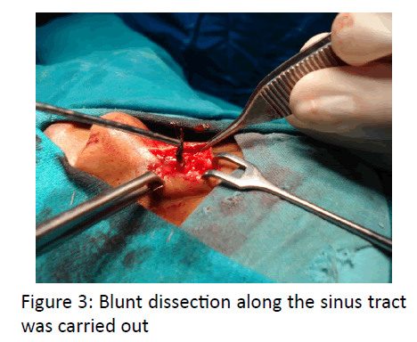
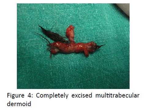
The child was taken for surgical excision of the dermoid under general anesthesia. An elliptical incision around the tuft of hair was made and blunt dissection along the sinus tract was done (Figures 2 and 3). A multitrabecular sinus was identified. The first tract ended blindly over the frontal bone at the glabella, the second tract extended from the mid portion of the first to end over the nasal bone. The third tract was a fistulous connection between the sinus tract to the superficial skin at the medial canthus of the right eye. None of the tracts showed any bony erosion on the underlying bones. All the tracts were dissected and the wound was sutured in layers. The histopathological report confirmed the diagnosis of dermoid (Figure 4). All sutures were removed on the 7th post-operative day and an aesthetic surgical scar was achieved. Hospital stay was uneventful.
Discussion
The Dermoid sinus with or without cyst is an extensive lesion extending into the nasal cartilage and bone, usually passing deeply at the osteocartilaginous junction on the dorsum of the nose.
The simplest cleft malformation in the front basal region is the dermal sinus which may be accompanied by dermoid cyst. This group includes a large proportion of the congenital nasal fistulas and dermoid cysts of the nose and frontal area, which are infrequently seen by the ENT surgeon. These fistulae may extend from the skin to the frontal bone leading to pressure atrophy and narrowing of the frontal sinus, if it is developed. They may extend through the skull base to the cranial cavity ending as an extradural cyst. Nasal fistulae can also pass through the dura ending in to a sub arachnoid or intradural cyst. The external opening in the skin may be inconspicuous and masked by a pigmented spot, so the condition remains undiagnosed until a subcutaneous fistulitis supervenes, possibly accompanied by intracranial complication, eg. Meningitis or encephalitis. The fistula opening is most commonly located on the nasal dorsum, rarely at the columella. With a sub dural epidermoid cyst, a supervening inflammatory complication can produce symptoms of meningitis, brain abscess or brain tumor; the possibility of such an anomaly should be considered whenever unexplained meningitis appears.
Their occurrence is due to congenital developmental anomaly and most accepted theory is prenasal theory. This theory suggests that neuroectodermal tract recedes, dermal attachments can follow its course. As the dura mater recedes from the prenasal space, it may pull the nasal ectoderm upward and inward to form a sinus or cysts [8].
Dermoid cyst is a relatively common congenital abnormality. It is a median nasal lesion found on the glabella, dorsum or tip of the nose between the alar cartilages or on the columella. It may be superficial or communicate with the deeper component through a tract between the nasal bones which may extend intracranial through the cribriform plate.
Preoperative radiological investigation of all cases of nasal dermoids should be done to diagnose any intracranial extension and to guide surgical plans.
CT and MRI have become the “gold standard” in radiologic evaluation of nasal dermoids.
CT and MRI provide complementary information and displace bony anatomy, soft tissue characterestic and CNS connections [9] respectively. The use of contrast differentiates between avascular dermoids and enhancing lesion such as haemangiomas and vascular teratomas.
Complete excision of dermoid cyst is treatment of choice. Surgical approaches for the removal of nasal dermoid depend upon extension of lesion. Different approaches have being advocated for removal of nasal dermoid which ranges from external approaches such as midline vertical incision, transverse incision, lateral rhinotomy, external rhinotomy, inverted U-incision, degloving procedure and craniotomy for intracranial extension of nasal dermoid.
Dermoid cyst is rare and often presents from birth appearing on the nose as a midline mass anywhere from the glabella to the columella. They rarely exceed 4 cm in size and possibly originate from inclusion of epidermal tissues in aberrant sites during embryonic development. A presumed dermoid cyst over the nasal root should be treated cautiously as this may represent a nasal glioma or a meningoencephalocoele with potential communication to the cranial cavity. Imaging and needle aspiration of the mass are necessary to confirm the source of the lesion before proceeding to excision.
References
- Denoyela F (1997) Nasal dermoid sinus cyst in children. Laryngoscope 107: 795-800.
- Rohrich RJ, Lwe JB and schwart MR (1999)The role of open rhinoplasty in management of nasal dermoid cysts. PlastreconstrSurg104:1459-1466.
- Kelly JH, Strome M and Hall B (1982) Surgical update on nasal dermoid. Arch otolaryngology 108: 239-242.
- Hughes (1980)The management of the congenital midline mass- a review. Otolaryngology-head and neck surgery 2:222-223.
- Jaffe BF(1981) Classification and management of anomalies of the nose. Otolaryngologic clinic of North America 14:989-1004.
- Wardinsky(1991) Nasal dermoid sinus cysts: association with intracranial extension and multiple malformations. Cleft palate palatecraniofac J 28:87-95.
- Reza Rahbar (2003)the presentation and management of nasal dermoid .Arch Otolaryngology head neck surg129:464-471.
- Pratt LW (1965) Midline cysts of the nasal dorsum: embryologic origin and treatment. Laryngoscope 75:968-980.
- Michelle Wyatt. Nasal obstruction in children. Scott Brown’s otolaryngology. Seventh Edn., chap 82: 1073-1074.


