Research Article - Biomedical Research (2017) Volume 28, Issue 9
A computation study on the effect of orientation of fibular allograft with screw in treating the femoral head necrosis
Zhenqiu Chen1, Xu Chen1*, Zhinan Hong2, Jianfa Chen1 and Guoju Hong21The First Affiliated Hospital, Guangzhou University of Chinese Medicine, Guangzhou, China
2Guangzhou University of Chinese Medicine, Guangzhou, China
- *Corresponding Author:
- Xu Chen
The First Affiliated Hospital
Guangzhou University of Chinese Medicinem China
Accepted on January 27, 2017
Abstract
Key steps for femoral head necrosis should meet the criteria of lesion debridement with less trauma and providing biology repair materials and structure support. To our knowledge, Fibular Allograft with Impaction Bone Grafting (FAIBG) as a minimally invasive technique could solve these problems, which has attained stable clinical effects. However, clinical doctors and researchers still debate the orientation of the implant. This study used computational biomechanical technique to explore the optimal orientation of FAIBG though investigating and analysing the stress distributions of anterolateral column and peak stress. The results indicated that the orientation of core decompression with FAIBG may attain better biomechanical conditions for the repair of osteonecrosis.
Keywords
Finite element analysis, Anterolateral column, Stress, Femoral head necrosis, Fibular allograft, Impaction bone graft.
Introduction
The common objective of every hip preservation procedure is to reserve the self-head of the patient with Femoral Head Necrosis (FHN) [1]. Several procedures, such as Core Decompression (CD), core decompression with fibular autograft, fan-shape decompression with fibula allograft, allogeneic fibular implantation and tantalum insertion have been developed to arrest the progression of osteonecrosis to avoid Femoral Head Collapse (FHC) and Total Hip Replacement (THR) in the early stage of FHN [2-14]. Key steps for FHN should meet the criteria of lesion debridement with less trauma and providing biology repair materials and structure support. To our knowledge, isolated CD shows limited success in delay the progression of FHC because it could not recover the biomechanical environment [15,16]. Autologous bone graft is often associated with serious trauma, high infection and longer recovery time [17]. Fibular Allograft with Impaction Bone Grafting (FAIBG) is a minimally invasive technique for preventing the mechanical failure of femoral head and interrupting the disease of FHC and THA, which attained stable clinical effects. However, clinical doctors and researchers still debate the path of the implant.
In this study, computational biomechanical technique was used to explore the optimal path of FAIBG though computing and analysing the stress distributions of anterolateral column and peak stress, which provides theoretical proof for clinical practice.
Materials and Methods
Generation of 3D geometry
CT image of the entire hip were acquired from an osteonecrotic patient without FHC. The resolution of the CT images was 1024 × 1024 pixels. The slice thickness was 0.1 mm. The images of DICOM format were input into the interactive medical image control system (mimics 15.1) to extract the 3D geometry of hip bone. The acetabular and femoral head cartilages were reconstructed based on anatomical data in the reverse engineering software Rapidform XOR3.
Parametric modelling
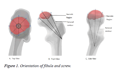
The parametric analysis was designed to explore the optimal path of FAIBG. The orientations of fibula with screw are schematically shown in Figure 1. The screw was place parallel to the fibula. Screw thread embeds in the fibula upside and the tail presses the distal fibula. The fibular geometry was a 6 mm radius and 90 mm length cylinder. The apex of the fibula was 5 mm at a distance from the cortical bone of bearing weight region. There are nine models were created.
Mesh model and material model
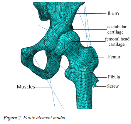
Tetrahedral elements of 4 mm size were used to generate mesh in ABAQUS (Figure 2). Constraints were applied to sacroiliac joint and pubic symphysis and load simulating single-legged stance was applied to the distal part of the femur. Five material properties were used to predict the biomechanical performance of hip joint [18-22]: Ecortical bone=15.1 GPa, Ecancellous bone=445 MPa, Elesion=124.6 Mpa, Ecartiage=10.5 MPa, Eti-6al-4v=113.8 GPa; vcortical bone=0.3, vcancellous bone=0.22, vlesion=0.152, vcartilage=0.45, vti-6al-4v=0.34. Friction coefficient between the femoral head cartilage and the acetabular cartilage was 0.01 and 0.42 between bone and stainless steel.
Results
Peak stress of bearing weight area
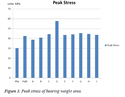
The peak stresses of bearing weight area were computed and shown in Figure 3. The peak stress in the physiologic control (Figure 3 (Phy)) was 30.25 Mpa. There are approximately 40.63% higher in pathological control (Figure 3 (Path)) than the physiologic level. After FAIBG treatment, the peak stresses fell off in A control and B control (Figures 3A and 3B) compared with the values obtained in the pathologic control, while in the other controls the peak stresses all rose.
Stress distribution of anteraloteral column
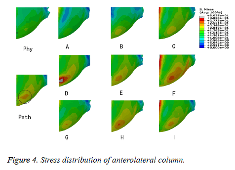
The stress distributions of anteraloteral column defined as an important indicator for FHC were plot in Figure 4. Figure 4 (Phy) showed that uniform stress distributed on anteraloteral column in physiologic control. A concentrated stress region appeared in pathologic control, which was shown in black circle. After FAIBG procedure, the stress significantly decreased and the concentrated stress regions disappeared in the A control (Figure 4A). The concentrated stress regions in the other controls still exist.
Discussion
Biomechanical reconstruction of the necrotic femoral head remains a complicated issue. Clinical doctors and researchers have developed various hip preservation procedures to protect the self-head of the FHN patient and used the finite element method to assess the curative effect thought analysing the biomechanical change of different materials [23]. These studies had shown that FEA has the ability to predict the collapse risk of femoral head with various surgical setting. However, relatively few FEM studies have considered the effect of orientation of FAIBG in Treating the FHN.
FAIBG has become the most potential substitute treatment for FHN, which combines the benefit of decompression with the added benefit of allogeneic repair and support materials integrated into the necrotic region. However, the optimal orientation of the implant has not been discriminated; hence, a study centered on the basic principles for FAIBG is still importance to avoid therapic failure and FHC. In this study, eleven models have been constructed and used to simulate nine orientation of the implant. The results showed that only the A control, as the orientation of core decompression, both reduce the peak stress and eliminate the concentrated stress region compared to the pathologic control. Hence, A control appears to be an optimal orientation of FAIBG in treating FHN.
Conclusion
In conclusion, parametric FEA is a useful tool for providing a biomechanical basis for clinical practice. The simulated results indicated that the orientation of core decompression for FAIBG may attain better biomechanical conditions for the repair of osteonecrosis.
Acknowledgements
There are no financial and personal relationships with other people or organizations that could inappropriately influence our work for all authors of this paper.
Conflict of Interests Statement
The authors declared no potential conflicts of interest with respect to the research, authorship, and/or publication of this article.
References
- Hungerford DS. Treatment of osteonecrosis of the femoral head: everythings new. J Arthroplasty 2007; 22: 91-94.
- Hungerford DS, Jones LC. Core decompression. Technique in Orthopaedics 2008; 23: 26-34.
- McGrory BJ, York SC, Iorio R, Macaulay W, Pelker RR. Current practices of AAHKS members in the treatment of adult osteonecrosis of the femoral head. J Bone Joint Surg Am 2007; 89: 1194-1204.
- Urbaniak JR, Harvey EJ. Revascularization of the femoral head in osteonecrosis. J Am AcadOrthopSurg 1998; 6: 44-54.
- Plakseychuk AY, Kim SY, Park BC, Varitimidis SE, Rubash HE. Vascularized compared with nonvascularized fibular grafting for the treatment of osteonecrosis of the femoral head. J Bone Joint Surg Am 2003; 85-85A: 589-96.
- Soucacos PN, Beris AE, Malizos K, Koropilias A, Zalavras H. Treatment of avascular necrosis of the femoral head with vascularized fibular transplant. ClinOrthopRelat Res 2001; 120-130.
- Buckley PD, Gearen PF, Petty RW. Structural bone-grafting for early atraumatic avascular necrosis of the femoral head. J Bone Joint Surg Am 1991; 73: 1357-1364.
- Keizer SB, Kock NB, Dijkstra PD, Taminiau AH, Nelissen RG. Treatment of avascular necrosis of the hip by a non-vascularised cortical graft. J Bone Joint Surg Br 2006; 88: 460-466.
- Guo X, Dou B, Zhou Y, Li Y. Surgical treatment of necrosis of the femoral head in early stages with core depression and allo-fibular grafting. ZhongguoXiu Fu Chong JianWaiKeZaZhi 2005; 19: 697-699.
- Bobyn JD, Poggie RA, Krygier JJ, Lewallen DG, Hanssen AD. Clinical validation of a structural porous tantalum biomaterial for adult reconstruction. J Bone Joint Surg Am 2004; 86-86A: 123-129.
- Xu WH, Yang SH, Li BX, Yang C, Ye SN, Tang X, Ye ZW. Allogeneic cortical bone cage support combining with autologous cancellous bone grafting for management femoral headnecrosis. Chin J ReparReconstrSurg 2009; 23: 527.
- Shi FL, Chen J, Li XH, Lu FX. Fan-shaped decompression and allograft fibula supporting internal fixation for treatment of early femoral head necrosis in adults. Chin J Tissue Eng Res 2013; 17: 7758.
- Yao H, Hu WH, Li HJ, Liu SY. Allogeneic fibular implantation for the treatment of femoral head necrosis: Clinical observation of 132 hips during 2.5 years follow-up. Chin J Tissue Eng Res 2013; 17: 3311.
- Lin ZJ, Su PJ, Wu ZQ, Gao DW, Li ZQ. Pith decompression of the femoral head and allograft fibula grafting for treatment of avascular necrosis of femoral head. ZhongguoGu Shang 2009; 22: 628-630.
- Camp JF, Colwell CW. Core decompression of the femoral head for osteonecrosis. J Bone Joint Surg Am 1986; 68: 1313-1319.
- Koo KH, Kim R, Ko GH, Song HR, Jeong ST. Preventing collapse in early osteonecrosis of the femoral head. A randomised clinical trial of core decompression. J Bone Joint Surg Br 1995; 77: 870-874.
- Veillette CJ, Mehdian H, Schemitsch EH, McKee MD. Survivorship analysis and radiographic outcome following tantalum rod insertion for osteonecrosis of the femoral head. J Bone Joint Surg Am 2006; 88: 48-55.
- Bae JY, Kwak DS, Park KS, Jeon I. Finite element analysis of the multiple drilling technique for early osteonecrosis of the femoral head. Ann Biomed Eng 2013; 41: 2528-2537.
- Brown TD, Hild GL. Pre-collapse stress redistributions in femoral head osteonecrosis--a three-dimensional finite element analysis. J BiomechEng 1983; 105: 171-176.
- Brown TD, Way ME, Ferguson AB. Mechanical characteristics of bone in femoral capital aseptic necrosis. ClinOrthopRelat Res 1981; 240-247.
- Grecu D, Pucalev I, Negru M, Tarnita DN, Ionovici N, Dita R. Numerical simulations of the 3D virtual model of the human hip joint, using finite element method, Rom J MorpholEmbryol 2010; 51: 151-155.
- Goffin JM, Pankaj P, Simpson AH. A computational study on the effect of fracture intrusion distance in three- and four-part trochanteric fractures treated with gamma nail and sliding hip screw. J Orthop Res 2014; 32: 39-45.
- Lee MS, Tai CL, Senan V, Shih C, Lo SW, Chen WP. The effect of necrotic lesion size and rotational degree on the stress reduction in transtrochaniteric rotational osteotomy for femoral head osteonecrosis- a three-dimensional finite-element simulation. ClinBiomech 2006; 21: 969-976.


