Case Report - Biomedical Research (2017) Volume 28, Issue 7
A case report of colon carcinoma featuring exophytic cavity growth
Chaohua Li1, Guangrui Shao1*, Yong Zhou2, Fengguo Sun1 and Yong Zhao11Department of Imaging, the Second Affiliated Hospital of Shandong University, China
2Department of General Surgery, the Second Affiliated Hospital of Shandong University, China
- *Corresponding Author:
- Guangrui Shao
Department of Imaging
The Second Affiliated Hospital of Shandong University
China
Accepted date: November 26, 2016
Abstract
We reported a case report of mass-like cecum and ascending colon carcinoma featuring exophytic cavity growth. The external lump size was about 84 mm × 72 mm that violated right-side illiopsoas and psoas major muscles. Preoperative CT diagnosis was performed to confirm a retroperitoneal cancer involving right hemicolon, psoas major and casiliopsoas muscles. Mesenchymoma was the paramount consideration. Besides, right hemicolon excision and involved psoas major excision were performed during operation. The postoperation pathology showed that colon cancer has involved iliopsoas and psoas major muscles. Above all, the patients recovered well after operation.
Keywords
Colon carcinoma, Exophytic growth, Right hemicolectomy
Introduction
Colon cancer is one of the most malignant tumours in the word and occurs frequently from age 40 to 70. In China, the rate of colon cancer is 13% [1]. There were few reports on colon cancer featuring exophytic growth, which was easily misdiagnosed as mesenchymoma. The best therapies for colon cancer were excision partly or totally. The lack of specificity, unapparent intestinal obstruction and late clarified diagnosis made poor prognosis.
Case History
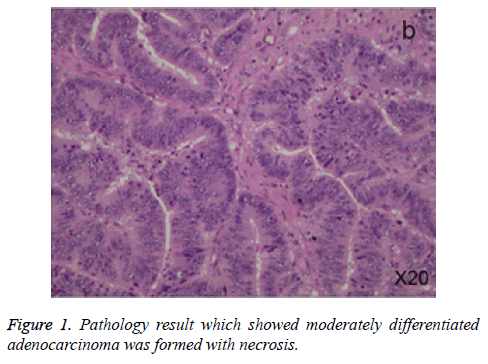
We reported a case report of mass-like cecum and ascending colon carcinoma featuring exophytic cavity growth here. A 39 year old male patient was admitted to hospital at February with right low abdominal pain. No constipation and hematochezia symptoms were observed. Preoperative physical examination showed palpable mass at low right abdomen. Moreover, CT examination also showed low right abdomen retroperitoneal heterogeneity-reinforced soft tissue mass with the size of 84 mm × 72 mm. Multiple liquidation necrosis was observed in the mass. Besides, there’s no apparent dividing line with adjacent psoas major muscle, iliopsoas muscle, cecum and colon ascendens intestinal walls. Balanced crassation of colon and cecum was observed. Multiple big lymph glands were found around the tumor, while obvious heterogenity reinforcement was visible under enhancement scan. CT diagnosis showed retroperitoneal cancer formation involving right hemicolon, right psoas major muscle and iliopsoas. Additionally, coloscope showed nodular lump was formed at colon ascendens or hepatic flexure, involving annulus area. The surface of the lump was irregular with multiple erosions. Enteric cavity showed obvious stenosis, making it hard for coloscope to go through. It is showed by pathology (Figure 1) that moderately differentiated adenocarcinoma was formed with necrosis. Swelling lymph gland was observed at the root of mesentery during operation, and the tumor lied in colon ascendens with the size of 5 cm × 3 cm × 3 cm. The tumor is hard that infiltrated towards right rear to retroperitoneum. Then a fixed hard lump was seen that was 15 cm × 15 cm × 10 cm size. The mass was adhered to right side external iliac blood vessel and ureter, involving right side iliopsoas and muscular fasciae of psoas. Right hemicolectomy was performed during operation. General pathology showed that right hemicolon moderately differentiated adenocarcinoma, soaking basic layer and adventitia. Luckily, no carcinoma was seen at lymph gland around the intestines. At last, right hemicolectomy and psoas major ablation were performed, making good recovery for postoperative patients.
Discussion
Colon invasive carcinoma could arise in exophytic or endophytic growth. However, most cases were endophytic and arose at left hemicolon, while few exophytic types were reported. A case report of right hemicolon cancer was reported here, featuring external cavity giant mass in retroperitoneum. Colon cancer usually occurred at 50 to 60 year old patients, and manifest asymmetry thickening in intestinal canal wall. Obvious enhancement could be seen under enhancement scan, with the tendency towards local and distant transfer. Electronic fiber coloscope and pathology could give a clarified diagnosis. It’s hard to distinguish from colon lymphoma and retroperitoneal malignant mesencymoma when colon cancer grew externally. Mesenchymoma occurred when the lesion of gastrointestinal tract grew externally. Moreover, malignant mesenchymoma usually occurred around 60 years old [2], while children and young people remained scarce [3,4]. Lobulated and irregular mass growing externally or internally could be seen on CT. The border was clear or obscure, and surrounding tissue was changed by pressure elapse. Obvious reinforcement could be seen under arterial or venous phase of enhancement scan, especially in portal vein phase. Necrosis and bleeding area had no obvious enhancement in mass center. Besides, no obvious decline was seen in equilibrium phase while the degree of enhancement was lower in delayed phase. Obvious blood vessel shadow and feeding artery could be seen in tumor at arterial phase. The diagnostic criteria of GIST was weakly positive or negative for immunohistochemical CD117 (+), CD34 (+), S-100 (-), Desmin and SMA [5].
Colon lymphoma had a lower morbidity than colon cancer and usually occurred at cecum. The onset age was younger than colon cancer. Intestinal wall incrassation was most commonly seen, while rag and surrounding infiltration symptom was rarely observed. Anabrosis and fistulous tract could be formed when growing externally. Colon, liver, spleen and intestine were most common focus for fistulous tract. Besides, light to moderate reinforcement was observed for lump under enhancement scan. However, reinforcement of colon cancer intensified obviously. Abdominopelvic cavity and retroperitoneal lymph node enlargement were usually seen in lymphoma, while only small lymph gland could be seen in colon cancer. The lymph gland in colon cancer rarely mixed together and usually occurred in regional lymph gland. The lymph gland of the patient reported herein was localized around mesentery with small size, which was consistent to the description.
Acknowledgement
This work was supported by Youth Funding of the Second Affiliated Hospital of Shandong University (Y2013010060).
References
- Siegel RL, Miller KD, Jemal A. Cancer statistics, 2016. CA Cancer J Clin 2016; 62: 10-29.
- Dematteo RP, Lewis JJ, Leung D, Mudan SS, Woodruff JM, Brennan MF. Two hundred gastrointestinal stromal tumors: recurrence patterns and prognostic factors for survival. Ann Surg 2000; 231: 51.
- Prakash S, Sarran L, Socci N, Dematteo RP, Eisenstat J, Greco AM. Gastrointestinal stromal tumors in children and young adults: a clinicopathologic, molecular, and genomic study of 15 cases and review of the literature. J Pediatr Hematol Oncol 2005; 27: 179-187.
- Miettinen M, Lasota J, Sobin LH. Gastrointestinal stromal tumors of the stomach in children and young adults: a clinicopathologic, immunohistochemical, and molecular genetic study of 44 cases with long-term follow-up and review of the literature. Am J Surg Pathol 2005; 29: 1373-1381.
- Miettinen M, Sobin LH, Lasota J. Gastrointestinal stromal tumors of the stomach: a clinicopathologic, immunohistochemical, and molecular genetic study of 1765 cases with long-term follow-up. Am J Surg Pathol 2005; 29: 52-68.
